Volume 8, Number 12—December 2002
Research
Induction of Inflammation by West Nile virus Capsid through the Caspase-9 Apoptotic Pathway
Figure 1

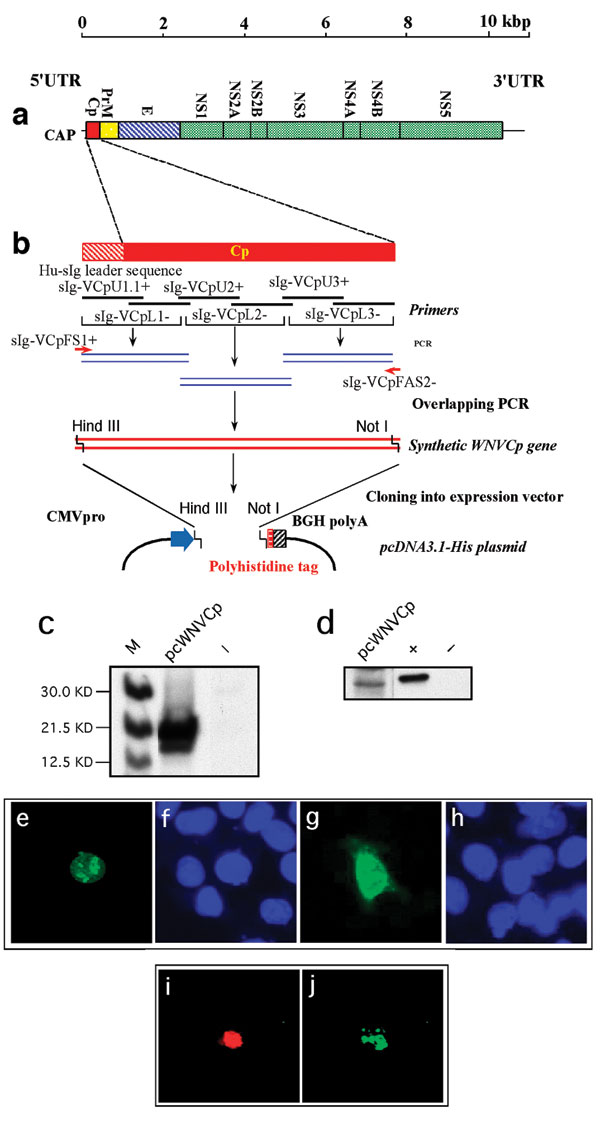
Figure 1. West Nile virus capsid (WNV-cp)-DJY protein expression induces apoptosis. Nuclear condensation was observed in HeLa cells transfected with pcWNV-Cp-DJY (a), a positive control pBax (b), or a negative control, pcDNA3.1 (c), under a 4,6-diamidino-2-phenylidole (DAPI) filter (magnification: 200X). Light microscopic observation on pcWNV-Cp-DJY (d) or pcDNA3.1 (e) plasmid transfected RD cells were examined in semithin sections stained with toluidine blue (magnification for d and e: 400X). Ultramicroscopic image of apoptotic cells were photographed from pcWNV-Cp-DJY transfected RD cells (f). DNA fragmentation in WNV capsid–expressing cell lines was examined by terminal desoxyriboxyl-desoxyriboxyl trasferase–mediated DVTP nick-end labeling (TUNEL) assay in HeLa (g), HEK 293 (i), and RD cells (k), and compared with DNA fragmentation from pcDNA3.1-transfected HeLa cells (m). Nuclear staining in HeLa (h), HEK 293 (j), and RD cells (l) were observed by using a DAPI filter and compared with control HeLa cells (n) (magnification for g through n: 400X). Cell membrane morphology changes were examined by annexin V staining/flow cytometry by using HeLa cells transfected with pcWNV-Cp-DJY or control pcDNA3.1 plasmids (o). The human neuroblastoma cell line SH-SY5Y was transfected with Bax as a positive control (p), pcWNV-Cp (r), or control plasmid (t) and examined by TUNEL assay. To visualize nuclear staining, cells transfected with pBax, pcWNV-Cp-DJY (q and s, respectively) or pcDNA3.1 (u) were stained with DAPI and observed using appropriate filters (magnification: 400X).