Skip directly to site content Skip directly to page options Skip directly to A-Z link Skip directly to A-Z link Skip directly to A-Z link
Volume 8, Number 12—December 2002
Research

Induction of Inflammation by West Nile virus Capsid through the Caspase-9 Apoptotic Pathway

Joo-Sung Yang*1, Mathura P. Ramanathan*1, Karuppiah Muthumani*, Andrew Y. Choo*, Sung-Ha Jin*, Qian-Chun Yu*, Daniel S. Hwang*, Daniel K. Choo*, Mark D. Lee*, Kesen Dang*, J. Joseph Kim†, David B. Weiner*Comments to Author , and WaixingTang
Author affiliations: *University of Pennsylvania, Philadelphia, Pennsylvania, USA; †Viral Genomix, Inc., Philadelphia, Pennsylvania, USA;

Main Article

Figure 2

Construction and subcellular expression of West Nile virus (WNV)–NY1999 capsid (Cp) gene–expressing plasmid, pcWNV-Cp-DJY: a, Genomic organization of WNV-NY1999 (10,945 bp) is outlined based on the published (GenBank accession no. AF202541). b, Cloning strategy for WNVCp gene–expressing plasmid, pcWNV-Cp-DJY. c, In vitro translated and immunoprecipitated 35S-labeled WNV-Cp visualized by SDS-PAGE. WNV-Cp–specific protein synthesis was compared to control generated by the vector backbone pcDNA3.1 (-). d, Protein expression by Western blot analysis, of WNV-Cp expression in HeLa cells. Subcellular location of WNV-Cp protein, in HeLa cells transfected with pcWNV-CpWT (e,f) or pcWNV-Cp-DJY (g,h) plasmids. 16 h posttransfection, the cells were visualized by indirect immunofluorescence. Typical nuclear staining was observed with the cells expressing WNV-CpWT (e) compared to the cells expressing WNV-Cp-DJY (g). TUNEL assay on the WNV-Cp–transfected cells, indicating nuclear condensation (i) due to expression of capsid (j).

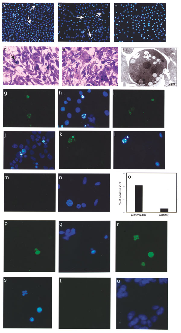
Figure 2. Construction and subcellular expression of West Nile virus (WNV)–NY1999 capsid (Cp) gene–expressing plasmid, pcWNV-Cp-DJY: a, Genomic organization of WNV-NY1999 (10,945 bp) is outlined based on the published (GenBank accession no. AF202541). b, Cloning strategy for WNVCp gene–expressing plasmid, pcWNV-Cp-DJY. c, In vitro translated and immunoprecipitated 35S-labeled WNV-Cp visualized by SDS-PAGE. WNV-Cp–specific protein synthesis was compared to control generated by the vector backbone pcDNA3.1 (-). d, Protein expression by Western blot analysis, of WNV-Cp expression in HeLa cells. Subcellular location of WNV-Cp protein, in HeLa cells transfected with pcWNV-CpWT (e,f) or pcWNV-Cp-DJY (g,h) plasmids. 16 h posttransfection, the cells were visualized by indirect immunofluorescence. Typical nuclear staining was observed with the cells expressing WNV-CpWT (e) compared to the cells expressing WNV-Cp-DJY (g). TUNEL assay on the WNV-Cp–transfected cells, indicating nuclear condensation (i) due to expression of capsid (j).

Main Article

1 These authors contributed equally.

Page created: July 19, 2010
Page updated: July 19, 2010
Page reviewed: July 19, 2010
The conclusions, findings, and opinions expressed by authors contributing to this journal do not necessarily reflect the official position of the U.S. Department of Health and Human Services, the Public Health Service, the Centers for Disease Control and Prevention, or the authors' affiliated institutions. Use of trade names is for identification only and does not imply endorsement by any of the groups named above.
file_external