Skip directly to site content Skip directly to page options Skip directly to A-Z link Skip directly to A-Z link Skip directly to A-Z link
Volume 8, Number 3—March 2002
Research

Eastern Equine Encephalomyelitis Virus Infection in a Horse from California

Robert P. Franklin*Comments to Author , Hailu Kinde†, Michele T. Jay‡, Laura D. Kramer†, Emily-Gene N. Green†, Robert E. Chiles†, Eileen Ostlund§, Stan Husted‡, Jonathan Smith¶, and Michael D. Parker¶
Author affiliations: *Humphrey, Giacopuzzi & Associates Equine Hospital, Somis, California, USA †University of California Davis, Davis, California, USA; ‡California Department of Health Services, Sacramento, California, USA; §National Veterinary Service Laboratory, Ames, Iowa, USA; ¶U.S. Army Medical Research Institute of Infectious Diseases, Fort Detrick, Maryland, USA;

Main Article

Figure 1

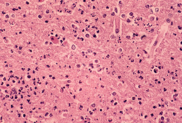
Photomicrograph of a section of the cerebral cortex from horse with Eastern equine encephalomyelitis virus infection. Note the dense neutrophilic response, vascular damage, and fibrin thrombi. Hematoxylin and eosine stain.

Figure 1. Photomicrograph of a section of the cerebral cortex from horse with Eastern equine encephalomyelitis virus infection. Note the dense neutrophilic response, vascular damage, and fibrin thrombi. Hematoxylin and eosine stain.

Main Article

Page created: July 15, 2010
Page updated: July 15, 2010
Page reviewed: July 15, 2010
The conclusions, findings, and opinions expressed by authors contributing to this journal do not necessarily reflect the official position of the U.S. Department of Health and Human Services, the Public Health Service, the Centers for Disease Control and Prevention, or the authors' affiliated institutions. Use of trade names is for identification only and does not imply endorsement by any of the groups named above.
file_external