Volume 9, Number 10—October 2003
Research
Escherichia coli O157 Exposure in Wyoming and Seattle: Serologic Evidence of Rural Risk
Cite This Article
Citation for Media
Abstract
We tested the hypothesis that rural populations have increased exposure to Escherichia coli O157:H7. We measured circulating antibodies against the O157 lipopolysaccharide in rural Wyoming residents and in blood donors from Casper, Wyoming, and Seattle, Washington, by enzyme immunoassay (EIA). EIA readings were compared by analysis of variance and the least squares difference multiple comparison procedure. Rural Wyoming residents had higher antibody levels to O157 LPS than did Casper donors, who, in turn, had higher levels than did Seattle donors (respective least squares means: 0.356, 0.328, and 0.310; p<0.05, Seattle vs. Casper, p<0.001, rural Wyoming vs. either city). Lower age was significantly correlated with EIA scores; gender; and, in rural Wyoming, history of bloody diarrhea, town, duration of residence, and use of nontreated water at home were not significantly correlated. These data suggest that rural populations are more exposed to E. coli O157:H7 than urban populations.
Escherichia coli O157:H7 is an important human pathogen. This organism can affect humans in a variety of ways, ranging from asymptomatic carriage (1) to diarrhea, bloody diarrhea (the most common manifestation of illness in culture-proven cases), and the postdiarrheal thrombotic microangiopathy, hemolytic uremic syndrome (HUS) (2). Infections with E. coli O157:H7 in the Pacific Northwest of the United States have been endemic (3) and epidemic (4). Vehicles transmitting this pathogen include unpasteurized milk and juice (5,6), undercooked beef (7), drinking water (8), and contact with infected persons (9).
Data from the Centers for Disease Control and Prevention (CDC) demonstrate higher incidences of E. coli O157:H7 infections in rural counties in the United States than urban (Paul Mead, unpub. data). Worldwide, rural populations have been postulated to be at greater risk for exposure to E. coli O157:H7 by virtue of increased exposure to animals or their excreta in Scotland (10,11); dairy farm visits have been implicated as a source for infection in Finland (12) and the United States (13); and animal contacts are a risk factor for the development of HUS in Switzerland (14). Serologic studies from Canada demonstrated higher frequencies of antibodies to the O157 lipopolysaccharide (LPS) side chain among residents of rural areas compared to residents of urban areas (15), and in Wisconsin children, manure and sheep contact were recently demonstrated to be risk factors for O157 seropositivity (16). Taken together, these data suggest more intense or more frequent human exposure to E. coli O157:H7 in nonurban areas.
Populations in the Pacific Northwest and Rocky Mountain states provide an opportunity to assess the frequency of exposure to E. coli O157:H7 through serologic studies. Antibodies to the O157 LPS follow natural infection with E. coli O157:H7 (17) and are believed to be quite specific (18) because they are rarely found in healthy people. Thus, circulating antibodies to the O157 LPS are potential markers of population exposure to E. coli O157:H7. We therefore attempted to assess the distribution of antibodies to this antigen in three different populations, encompassing a gradient of population density.
Study Participants
Participants were selected for inclusion in this study if they were >16 years of age, weighed >54 kg, and participated in voluntary cholesterol screening in several rural western Wyoming towns (population A), or donated blood to the Wyoming State (population B) or Puget Sound (population C) blood banks, and provided informed consent. The Institutional Review Boards of the Children’s Hospital and Regional Medical Center (Seattle, Washington) and the University of Wyoming (Laramie, Wyoming) approved this study before participants were enrolled.
Population A consisted of 485 residents of Star Valley, Wyoming. This valley has extensive agricultural land usage and consists of a series of small towns along U.S. Highway 89 in Lincoln County in the northwestern part of the state; town populations range from 100 to 1,200 residents. One of these towns had an E. coli O157:H7 outbreak in 1998 (19). During a local health fair conducted in the towns of Afton, Thayne, and Alpine during May 1999, participants donated 5 mL of blood during phlebotomy for screening to detect hyperlipidemia and answered a questionnaire regarding age, gender, treated versus nontreated domestic water supply, history of bloody diarrhea, location of sampling, and town of residence.
Population B was composed of 196 blood donors at United Blood Services (UBS), Casper, Natrona County, Wyoming. UBS, Wyoming’s only blood bank, obtains most of its blood from donors residing within Casper, the second largest municipality in the state (population 49,644 residents [20] in 52.8 km2 [Mike Jun, pers. comm.]). This site was chosen for study because of its presumed intermediate intensity of exposure to agriculture. Volunteers provided 5 mL of whole blood in a separate tube as part of the donation. Gender and age data were available for 127 (65%) of the participants.
Population C consisted of 104 blood donors at the Puget Sound Blood Center, Seattle, Washington, all of whom resided in urban or suburban Puget Sound municipalities (including Seattle and surrounding communities) and provided an additional 5 mL of blood for research. This group was presumed to have less agricultural exposure than populations A or B. Age and gender were recorded for all but two donors. Serum specimens from populations B and C were collected during the spring and summer of 1999.
Cattle and human density data for Lincoln, Natrona, and King Counties are provided in Table 1. These data demonstrate the rural-to-urban human population density and cattle-to-human ratios for the populations chosen. Blood samples were centrifuged within 3 hours of donation; serum samples were separated from the packed erythrocytes and stored at –70°C until assayed.
Detection of Antibodies to E. coli O157 LPS
E. coli O157:H7 LPS was purified from strain 86-24 (21) by using phenol extraction (22). Purified LPS underwent electrophoresis in a 12% polyacrylamide sodium dodecyl sulfate-polyacrylamide gel electrophoresis gel and then was transferred to a membrane and probed with antibodies to the LPS antigen (23). Serum specimens were screened for antibodies to the O157 LPS with an enzyme immunoassay (EIA) (24), modified by CDC. Briefly, in preliminary experiments, the optimal LPS concentrations for coating plates and diluting serum samples were determined by block titration with phosphate-buffered saline (PBS) (0.01 M, pH 7.2) and a known positive human sample. Optimal dilutions of 1:20 for serum and 1:160 of the antigen stock solution (corresponding to 200 μL of a resuspended pellet from a 50-mL overnight culture in Luria broth [25]) were demonstrated (data not shown).
Next, individual wells of an Immulon II plate (Dynex Technology, Franklin, MA) were coated with diluted antigen and incubated (4°C, overnight) to enable the target antigen to adhere to the plates. Then, 150 μL of PBS containing 1% fetal bovine serum and 0.5% nonfat dry milk (PBS-M-FBS) was placed in each well. The plates were then incubated in a blocking step (room temperature, 2 hours) to prevent nonspecific antibody adherence to the plates. Fluid was then removed, and the plates were washed four times with 0.01 M PBS containing 0.05% Tween 20 (PBS-T).
Ten microliters of diluted human serum sample was added to 190 μL of PBS-M-FBS containing 0.05% Tween 20 (PBS-T-M-FBS), placed into wells of the microtiter plates, and incubated (37°C, 1 h). The fluid was then removed, and the plates were washed four times with PBS-T. One hundred microliters of alkaline-phosphatase–labeled goat antihuman immunoglobulin (Ig) G/IgM/IgA (heavy and light chain) (Kierkegaard & Perry Laboratories, Inc., Gaithersburg, MD), diluted 1:10,000 in PBS-T-M-FBS, was then placed in each well and incubated (37°C, 1 h). The plates were then washed four times with PBS-T.
A substrate solution containing 97 mL diethanolamine, 0.2 g sodium azide, 100 mg magnesium chloride-6H2O, and 800 mL distilled water was adjusted to a pH of 9.8 with 1 M hydrochloric acid. One p-nitrophenyl phosphate tablet (Sigma Chemical Co., St. Louis, MO) was then added to 5 mL of substrate solution. One hundred microliters of this solution was placed in each well and incubated (room temperature, 25 min). The reaction was halted by adding 50 μL of 3 M sodium hydroxide to each well, and the optical densities of each well were read in a dual wavelength micro-EIA reader at λ = 405 nm with background correction at λ = 540 nm (Elx 800, Bio-Tek Instruments, Winooski, VT). Each serum sample was assayed in duplicate, and the values were averaged.
Serum from a patient with HUS caused by E. coli O157:H7 and serum from a study participant without known E. coli O157:H7 infection in population A were included as duplicates on each plate as positive and negative controls, respectively, and values were averaged. Each plate also contained controls without antigen or primary or secondary antibody. All plates were normalized linearly in relation to the positive control in the first group of serum samples tested.
Analysis
The complete dataset was first studied by analysis of variance (ANOVA, Proc GLM, SAS Institute, Inc., Cary, NC) in a model with EIA readings as the dependent variable, gender and town/city as class-independent variables, and age as a continuous independent variable. Initially, all interactions were included in the model, but interactions not contributing significantly to the model were dropped from subsequent analyses. Multiple comparisons were analyzed by using the protected Fisher least squares differences (LSD) test after confirming that the p value of the model as a whole was <0.05. The data were approximately normally distributed, as demonstrated by a Wilk-Shapiro statistic >0.98 (either for the dataset as a whole or for each region separately, Proc UNIVARIATE, SAS Institute, Inc.) and by visualization of the residuals plot. However, as assumptions of normal distribution of the data are difficult to confirm robustly, the data were also analyzed after transformation of these values into binary form with arbitrarily chosen cutpoints at the 80th and 90th percentiles of the EIA scores or with the entire range of EIA scores categorized at 0.05 increments, using stepwise logistic regression (Proc LOGISTIC, SAS Institute, Inc.) with the same independent variables as described above for the ANOVA (26). Statistically, p values <0.05 were considered significant for comparisons, and p values <0.05 were set as the criterion for entry and for retention into logistic regression models.
Figure
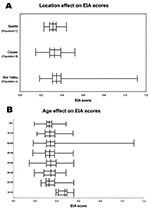
Figure. Box plot analysis for enzyme immunoassay (EIA) values, by populations. X axis represents EIA scores for study participants.Vertical line in each box represents the median for each population. The left and...
We tested 785 serum samples for antibody reactivity. The summary statistics for the O157 EIA are provided in the Figure (panel A), and the demographic characteristics of each population contributing these serum specimens are provided in Table 1. The average age of the study participants was significantly younger in populations B and C, than in population A. The populations did not differ significantly with respect to gender.
EIA scores were significantly related to both age (p<0.01) and population (p<0.001) in ANOVA. The effect of age is illustrated in the Figure (panel B). Age and gender information were not available for 35% of population B, but the distributions of EIA scores did not differ between those observations with and without these data, suggesting that the loss of data did not bias the results. The same two variables, age and population, also were significantly (p<0.05) associated with EIA score in logistic regression analysis, irrespective of whether the dependent variable modeled was the 80th percentile (Table 2) or the 90th percentile of the EIA score or increased increment of the EIA score (data not shown).
Within the rural Wyoming group, the data allowed analysis of O157 LPS EIA values’ association with additional variables, including the duration of residence in the area, occurrence of bloody diarrhea, use of a chlorinated water supply at the residence, and town of residence within Star Valley (Table 3). None of these variables was significantly associated with EIA mean values. Residents living in the area for <2 years tended to have higher average EIA values (0.380) than those who resided there for longer periods (2–5 years, 0.346; >5 years, 0.355).
These data suggest that rural residents have greater exposure to an antigen or antigens that produce antibodies to the E. coli O157 LPS antigen than do urban residents. However, we cannot state with certainty that the precipitating antigen was actually a pathogenic E. coli O157:H7. Because the O157 LPS antigen can be expressed by nonpathogenic E. coli (27), Citrobacter freundii (28), and E. hermanii (29), knowing the nature of the antigen during the immunizing event in the participants studied is not possible. Nonetheless, antibodies to the E. coli O157 LPS antigen plausibly represent exposure to pathogenic E. coli O157:H7, especially as examples exist of asymptomatic carriage of E. coli O157:H7 inducing an antibody response to O157 LPS (1,30). We believe, therefore, that this serologic reactivity likely represents actual exposure to pathogenic E. coli O157:H7. Also, our assay did not distinguish the classes of antibodies that were reactive in the EIA, so we cannot make estimates about the timing of the exposure based on class of antibody detected. However, IgA, IgG, and IgM antibodies to the O157 LPS are each ephemeral after natural symptomatic infections (31). Thus, the antibodies that we detected in this study quite likely represent recent exposure to the antigen.
The EIA levels were not proportional to cattle density per land area, a value that was similar in each of the three study sites. Thus, human exposure to E. coli O157:H7 cannot be attributed simply to cattle presence within counties. However, in rural counties, a higher proportion of residents might be involved in activities that bring them in contact with E. coli O157:H7, including animal contact. Our survey was not designed to measure such exposures within counties. Indeed, cattle-to-human spatial proximity in Ontario is a risk factor for infection in a novel application of livestock density indicators and disease incidence (32). Alternatively, rural residents might have a higher frequency of exposure to wild animals that carry E. coli O157:H7, such as deer (33), their excreta, or water that has been contaminated with their excreta. However, if a link between any animal source of E. coli O157:H7 and human exposure to this pathogen is to be further investigated, population distributions within counties, proximities of humans to animals and their excreta, and presence of E. coli O157:H7 in the environment will all need to be examined to determine the modes of contact.
Our data differ from results of previous studies in terms of statistical analysis and also because we included a gradient of population density. Specifically, our principal analysis did not require assignment of persons to seropositive or seronegative status categories. While some relation might exist between percentage of persons who are designated as having reactive or nonreactive status on the basis of cutoffs and the comparative distribution of serologic reactivities in populations, we believe that, when examining continuous variables, a test that compares values as continuous measurements (such as analysis of variance) provides more information and is less arbitrary than the assignment of categorical positives and negatives. We did, however, also examine proportions above two cutpoints, and the same trends were noted. For unknown reasons, we identified higher EIA scores in younger study participants, whereas in Ontario, IgG antibodies to the O157 LPS were highest in participants in their fifth decade of life (15). Comparing our age-related EIA scores to those reported recently from Wisconsin, where older children had higher seropositive rates, is not possible because the latter study focused on a child population and did not evaluate older Wisconsin residents (16).
We caution against interpreting our data to mean that rural populations are immune to, and thereby protected from, E. coli O157:H7 infections. Many study participants had antibody levels that were probably too low to confer protection, even if one assumes that antibodies to this antigen protect against infection. In this regard, an E. coli O157:H7 infection, which almost always induces a brisk and high-titer humoral immune response to the O157 LPS antigen, may not confer a permanently protective response, as evidenced by documentations of recurrent infections (34,35). However, in a recent E. coli O157:H7 outbreak in the Star Valley, a higher frequency of antibody levels to the O157 LPS among the resident population was proposed as the reason for a lower attack rate among residents than among visitors (19). We also urge against generalizing the trend observed in this study, which is derived from the analysis of only three populations, to all rural populations, without additional, more widespread, studies because the populations studied might not be representative. Nonetheless, our data are consistent with the hypothesis that rural residence carries with it a greater risk for exposure to E. coli O157:H7 than does urban residence.
In summary, we have identified an age-dependent, gender-independent, risk for probable exposure to E. coli O157:H7 in persons living in rural communities. This exposure frequency is plausibly environmental, rather than foodborne, in origin because food is distributed widely throughout North America. However, the possibility exists that particular food consumption practices, such as drinking raw milk in rural communities, as has been noted in the United Kingdom (36,37), might have been responsible for this exposure. We also cannot exclude the possibility that the differences observed relate to the nature of the participants studied. That is, donors to blood banks might have different exposures than participants in lipid screenings at health fairs. Future studies should attempt to identify the points of exposure to this antigen, confirm that E. coli O157:H7 is, indeed, the source of the inciting antigen, and, if it is, minimize human contact with this pathogen.
Dr. Haack is a resident at the Mayo Clinic, Rochester, Minnesota. This work was performed as a thesis project at the University of Washington School of Medicine.
Acknowledgments
We thank Jennifer L. Falkenhagen for assistance in manuscript preparation, Paul Mead for sharing unpublished data with us, and William Bibb for advice concerning enzyme immunoassay protocols.
The Escherichia coli Gift Fund at the Children’s Hospital and Regional Medical Center and NIH grant DK52081 support this project.
References
- Wilson JB, Clarke RC, Renwick SA, Rahn K, Johnson RP, Karmali MA, Vero cytotoxigenic Escherichia coli infection in dairy farm families. J Infect Dis. 1996;174:1021–7.PubMedGoogle Scholar
- Tarr PI. Escherichia coli O157:H7: clinical, diagnostic, and epidemiological aspects of human infection. Clin Infect Dis. 1995;20:1–8.PubMedGoogle Scholar
- MacDonald KL, O’Leary MJ, Cohen ML, Norris P, Wells JG, Noll E, Escherichia coli O157:H7, an emerging gastrointestinal pathogen. Results of a one-year, prospective, population-based study. JAMA. 1988;259:3567–70. DOIPubMedGoogle Scholar
- Bell BP, Goldoft M, Griffin PM, Davis MA, Gordon DC, Tarr PI, A multistate outbreak of Escherichia coli O157:H7–associated bloody diarrhea and hemolytic uremic syndrome from hamburgers. The Washington experience. JAMA. 1994;272:1349–53. DOIPubMedGoogle Scholar
- Keene WE, Hedberg K, Herriott DE, Hancock DD, McKay RW, Barrett TJ, A prolonged outbreak of Escherichia coli O157:H7 infections caused by commercially distributed raw milk. J Infect Dis. 1997;176:815–8. DOIPubMedGoogle Scholar
- Besser RE, Lett SM, Weber JT, Doyle MP, Barrett TJ, Wells JG, An outbreak of diarrhea and hemolytic uremic syndrome from Escherichia coli O157:H7 in fresh-pressed apple cider. JAMA. 1993;269:2217–20. DOIPubMedGoogle Scholar
- Riley LW, Remis RS, Helgerson SD, McGee HB, Wells JG, Davis BR, Hemorrhagic colitis associated with a rare Escherichia coli serotype. N Engl J Med. 1983;308:681–5. DOIPubMedGoogle Scholar
- Swerdlow DL, Woodruff BA, Brady RC, Greiffin PM, Tippen S, Donnell HD Jr, A waterborne outbreak in Missouri of Escherichia coli O157:H7 associated with bloody diarrhea and death. Ann Intern Med. 1992;117:812–9.PubMedGoogle Scholar
- Belongia EA, Osterholm MT, Soler JT, Ammend DA, Braun JE, MacDonald KL. Transmission of Escherichia coli 0157:H7 infection in Minnesota child day-care facilities. JAMA. 1993;269:883–8. DOIPubMedGoogle Scholar
- Locking ME, O’Brien SJ, Reilly WJ, Wright EM, Campbell DM, Coia JE, Risk factors for sporadic cases of Escherichia coli O157 infection: the importance of contact with animal excreta. Epidemiol Infect. 2001;127:215–20. DOIPubMedGoogle Scholar
- O’Brien SJ, Adak GK, Gilham C. Contact with farming environment as a major risk factor for Shiga toxin (Vero cytotoxin–producing Escherichia coli O157 infection in humans. Emerg Infect Dis. 2001;7:1049–51. DOIPubMedGoogle Scholar
- Lahti E, Eklund M, Ruutu P, Siitonen A, Rantala L, Nuorti P, Use of phenotyping and genotyping to verify transmission of Escherichia coli O157:H7 from dairy farms. Eur J Clin Microbiol Infect Dis. 2002;21:189–95. DOIPubMedGoogle Scholar
- Crump JA, Sulka AC, Langer AJ, Schaben C, Crielly AS, Gage R, An outbreak of Escherichia coli O157:H7 infections among visitors to a dairy farm. N Engl J Med. 2002;347:555–60. DOIPubMedGoogle Scholar
- Kernland KH, Laux-End R, Truttmann AC, Reymond D, Bianchetti MG. [How is hemolytic-uremic syndrome in childhood acquired in Switzerland?]. [In German]. Schweiz Med Wochenschr. 1997;127:1229–33.PubMedGoogle Scholar
- Reymond D, Johnson RP, Karmali MA, Petric M, Winkler M, Johnson S, Neutralizing antibodies to Escherichia coli Vero cytotoxin 1 and antibodies to O157 lipopolysaccharide in healthy farm family members and urban residents. J Clin Microbiol. 1996;34:2053–7.PubMedGoogle Scholar
- Belongia EA, Chyou PH, Greenlee RT, Perez-Perez G, Bibb WF, De Vries EO. Diarrhea incidence and farm-related risk factors for Escherichia coli 0157:H7 and Campylobacter jejuni antibodies among rural children. J Infect Dis. 2003;187:1460–8. DOIPubMedGoogle Scholar
- Banatvala N, Griffin PM, Greene KD, Barrett TJ, Bibb WF, Green JH, The United States National Prospective Hemolytic Uremic Syndrome Study: microbiologic, serologic, clinical, and epidemiologic findings. J Infect Dis. 2001;183:1063–70. DOIPubMedGoogle Scholar
- Reida P, Wolff M, Pohls HW, Kuhlmann W, Lehmacher A, Aleksic S, An outbreak due to enterohaemorrhagic Escherichia coli O157:H7 in a children day care centre characterized by person-to-person transmission and environmental contamination. Zentralbl Bakteriol. 1994;281:534–43.PubMedGoogle Scholar
- Olsen SJ, Miller G, Kennedy M, Higgins C, Walford J, McKee G, A waterborne outbreak of Escherichia coli O157:H7 infections and hemolytic uremic syndrome: implications for rural water systems. Emerg Infect Dis. 2002;8:370–5. DOIPubMedGoogle Scholar
- Profile of general demographic characteristics: 2000. Geographic area: Casper City, Wyoming. Available from: URL: http://www.census.gov/prod/cen2000/dp1/2kh56pdf
- Tarr PI, Neill MA, Clausen CR, Newland JW, Neill RJ, Moseley SL. Genotypic variation in pathogenic Escherichia coli O157:H7 isolated from patients in Washington, 1984–1987. J Infect Dis. 1989;159:344–7.PubMedGoogle Scholar
- Inzana TJ, Pichichero ME. Lipopolysaccharide subtypes of Haemophilus influenzae type b from an outbreak of invasive disease. J Clin Microbiol. 1984;20:145–50.PubMedGoogle Scholar
- Bilge SS, Vary JC Jr, Dowell SF, Tarr PI. Role of the Escherichia coli O157:H7 O side chain in adherence and analysis of an rfb locus. Infect Immun. 1996;64:4795–801.PubMedGoogle Scholar
- Toth I, Barrett TJ, Cohen ML, Rumschlag HS, Green JH, Wachsmuth IK. Enzyme-linked immunosorbent assay for products of the 60-megadalton plasmid of Escherichia coli serotype O157:H7. J Clin Microbiol. 1991;29:1016–9.PubMedGoogle Scholar
- Sambrook J, Fritsch EF, Maniatis T. Molecular cloning: a laboratory manual. 2nd ed. Cold Spring Harbor (NY): Cold Spring Harbor Laboratory Press; 1989.
- Glantz SS, Glantz SA. Primer of biostatistics. 4th ed. New York: McGraw-Hill Health Professions Division; 1997.
- Tarr PI, Schoening LM, Yea YL, Ward TR, Jelacic S, Whittam TS. Acquisition of the rfb-gnd cluster in evolution of Escherichia coli O55 and O157. J Bacteriol. 2000;182:6183–91. DOIPubMedGoogle Scholar
- Bettelheim KA, Evangelidis H, Pearce JL, Sowers E, Strockbine NA. Isolation of a Citrobacter freundii strain which carries the Escherichia coli O157 antigen. J Clin Microbiol. 1993;31:760–1.PubMedGoogle Scholar
- Chapman PA. Evaluation of commercial latex slide test for identifying Escherichia coli 0157. J Clin Pathol. 1989;42:1109–10. DOIPubMedGoogle Scholar
- Bielaszewska M, Janda J, Blahova K, Minarikova H, Jokova E, Karmali MA, Human Escherichia coli O157:H7 infection associated with the consumption of unpasteurized goat’s milk. Epidemiol Infect. 1997;119:299–305. DOIPubMedGoogle Scholar
- Ludwig K, Bitzan M, Bobrowski C, Muller-Wiefel DE. Escherichia coli O157 fails to induce a long-lasting lipopolysaccharide-specific, measurable humoral immune response in children with hemolytic-uremic syndrome. J Infect Dis. 2002;186:566–9. DOIPubMedGoogle Scholar
- Valcour JE, Michel P, McEwen SA, Wilson JB. Associations between indicators of livestock farming intensity and incidence of human shiga toxin–producing Escherichia coli infection. Emerg Infect Dis. 2002;8:252–7. DOIPubMedGoogle Scholar
- Rice DH, Hancock DD, Besser TE. Verotoxigenic E. coli O157 colonisation of wild deer and range cattle. Vet Rec. 1995;137:524. DOIPubMedGoogle Scholar
- Siegler RL, Griffin PM, Barrett TJ, Strockbine NA. Recurrent hemolytic uremic syndrome secondary to Escherichia coli O157:H7 infection. Pediatrics. 1993;91:666–8.PubMedGoogle Scholar
- Robson WL, Leung AK, Miller-Hughes DJ. Recurrent hemorrhagic colitis caused by Escherichia coli O157:H7. Pediatr Infect Dis J. 1993;12:699–701. DOIPubMedGoogle Scholar
- Sibbald CJ, Sharp JC. Campylobacter infection in urban and rural populations in Scotland. J Hyg (Lond). 1985;95:87–93. DOIPubMedGoogle Scholar
- Barrett NJ. Communicable disease associated with milk and dairy products in England and Wales: 1983–1984. J Infect. 1986;12:265–72. DOIPubMedGoogle Scholar
Figure
Tables
Cite This ArticleTable of Contents – Volume 9, Number 10—October 2003
| EID Search Options |
|---|
|
|
|
|
|
|
Please use the form below to submit correspondence to the authors or contact them at the following address:
P. I. Tarr, Edward Mallinckrodt Department of Pediatrics and Department of Molecular Microbiology, Washington University School of Medicine, and the Division of Gastroenterology, St. Louis Children's Hospital, St. Louis, MO 63110, USA; fax: 314-286-2911
Top