Volume 13, Number 9—September 2007
Dispatch
Equine Rhinosporidiosis in United Kingdom
Figure 1
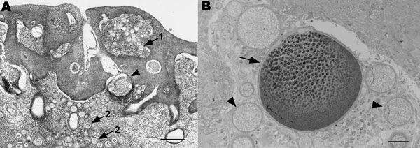
Figure 1. A) Section of nasal mucosa showing multifocal hyperplasia, juvenile sporangia (arrows) within the mucosal epithelium (1) and the lamina propria mucosae (2), and mature sporangia (arrowhead). A multifocal mixed inflammatory infiltrate can be seen within the mucosa. Stain, hematoxylin and eosin; magnification ×4; scale bar, 250 μm. B) Semithin section of nasal mucosa with juvenile sporangia (arrowheads) and a mature sporangium (arrow) with a lymphoplasmacellular inflammatory infiltrate within the lamina propria mucosae. Stain, toluidine blue; magnification ×10; scale bar, 40 μm.
1Current affiliation: The Royal Veterinary College, Hatfield, United Kingdom
2Current affiliation: Rest Associates, Swaffham Prior, Cambridge, United Kingdom
Page created: July 01, 2010
Page updated: July 01, 2010
Page reviewed: July 01, 2010
The conclusions, findings, and opinions expressed by authors contributing to this journal do not necessarily reflect the official position of the U.S. Department of Health and Human Services, the Public Health Service, the Centers for Disease Control and Prevention, or the authors' affiliated institutions. Use of trade names is for identification only and does not imply endorsement by any of the groups named above.