Volume 14, Number 1—January 2008
THEME ISSUE
International Polar Year
Dispatch
Human Ophthalmomyiasis Interna Caused by Hypoderma tarandi, Northern Canada
Philippe R.S. Lagacé-Wiens* , Ravi Dookeran*, Stuart Skinner*, Richard Leicht*, Douglas D. Colwell†, and Terry D. Galloway*
Figure 2
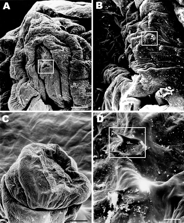
Figure 2. Scanning electron microscope images of the parasite from an 11-year-old Inuit boy, Nunavut, Canada. A) Anterior end of the maggot. The cephalic segment is evident; mouth and mouth hooks are present (boxed). Scale bar = 50 μm. B) The characteristic cephalic sensory array (boxed). Scale bar = 10 μm. C) Posterior segments of the maggot. Scale bar = 100 μm. D) Spiracular openings on the posterior segments of the maggot characteristic of first instar of Hypoderma. Scale bar = 10 μm.
Page created: July 08, 2010
Page updated: July 08, 2010
Page reviewed: July 08, 2010
The conclusions, findings, and opinions expressed by authors contributing to this journal do not necessarily reflect the official position of the U.S. Department of Health and Human Services, the Public Health Service, the Centers for Disease Control and Prevention, or the authors' affiliated institutions. Use of trade names is for identification only and does not imply endorsement by any of the groups named above.