Volume 14, Number 11—November 2008
Dispatch
Pyemotes ventricosus Dermatitis, Southeastern France
Figure 2

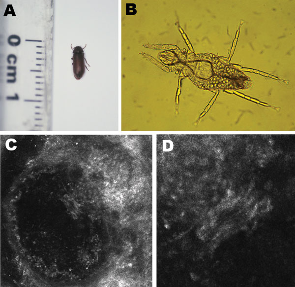
Figure 2. Organisms involved in transmission of Pyemotes ventricosus dermatitis. A) Common furniture beetle (Anobium punctatum) (5 × 2 mm). B) Nongravid female P. ventricosus mite (210 × 40 μm). C) Confocal laser scanning microscopy (CLSM) image (CLSM Vivascope 1500 microscope; Lucid Inc., Rochester, NY, USA) of a P. ventricosus mite (210 × 40 μm) in its microvesicle. D) Higher magnification of the microvesicle in panel C (light area in the center) (magnification ×4).
Page created: July 18, 2010
Page updated: July 18, 2010
Page reviewed: July 18, 2010
The conclusions, findings, and opinions expressed by authors contributing to this journal do not necessarily reflect the official position of the U.S. Department of Health and Human Services, the Public Health Service, the Centers for Disease Control and Prevention, or the authors' affiliated institutions. Use of trade names is for identification only and does not imply endorsement by any of the groups named above.