Volume 14, Number 12—December 2008
Dispatch
Transmission of Atypical Bovine Prions to Mice Transgenic for Human Prion Protein
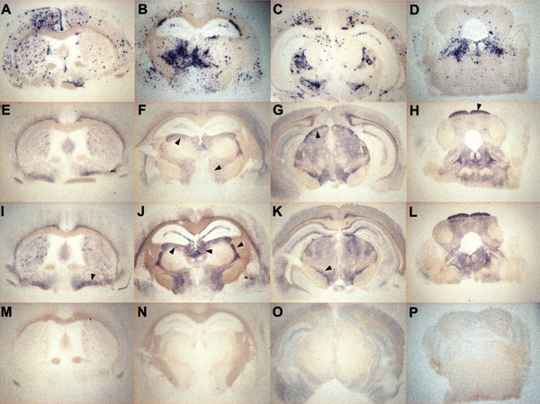
Figure 2

Figure 2. Representative histoblots in 4 different anteroposterior sections showing the distribution of disease-specific PrPres deposits in the brains of tg650 mice infected with bovine spongiform encephalopathy (BSE) or L-type BSE. Panels A–D show infection with BSE (second passage of France 3). Panels E–H show infection with L-type BSE (first passage of France 7). Panels I–L show infection with L-type BSE (second passage of Italy). Panels M–P show brain sections of an age-matched, mock-infected mouse, euthanized while healthy at 700 days postinfection, for comparison. Note the differing aspect and distribution of PrPres deposits between brain of mice infected with BSE and BSE-L (arrowheads). Assignment of the positive brain regions has been made according to a mouse brain atlas after digital acquisition.