Volume 14, Number 2—February 2008
Research
Genetic Characterization of Feline Leukemia Virus from Florida Panthers
Figure 1
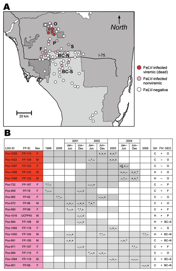
Figure 1. A) Prevalence and distribution of 19 Florida panthers, sampled 1999–2005, showing evidence of feline leukemia virus (FeLV) exposure. All antigen-positive panthers (red) are clustered in the Okaloacoochee Slough State Forest (O). PCR-positive and/or antibody-positive (pink) pumas were found there also, as well as in the surrounding areas including Florida Panther National Wildlife Refuge (F), private lands (P), Big Cypress Seminole Indian Reservation (S), and Big Cypress North and South (BC-N, BC-S respectively). All but 2 infected panthers were found north of Interstate 75. B) Information on affected panthers. Gray shading indicates timeline for monitoring of individual panthers until death. Symbols within gray boxes indicate presence (+), absence (–), or no data (*) for FeLV antigen in serum, FeLV sequence recovered by PCR, or presence of antibodies against FeLV in serum, respectively. FP-122 was antigen negative when tested 1 month previously (§). LGD ID, Laboratory of Genomic Diversity identification number; FP ID, Florida panther identification number; GH, genetic heritage; FIV, feline immunodeficiency virus; GEO, geographic locale; C, canonical (pure) Florida panther; H, Texas hybrid.