Volume 14, Number 6—June 2008
Dispatch
Nosocomial Outbreaks Caused by Leuconostoc mesenteroides subsp. mesenteroides
Germán Bou* , Jesús Luis Saleta*, Juan Antonio Sáez Nieto†, Mar Tomás*, Silvia Valdezate†, Dolores Sousa*, Francisco Lueiro*, Rosa Villanueva*, Maria Jose Pereira*, and Pedro Llinares*
Figure 2
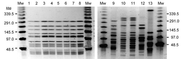
Figure 2. Band pattern obtained by pulsed-field gel electrophoresis of selected Leuconostoc mesenteroides subsp. mesenteroides (LM) isolates. Mw, molecular weight marker at indicated sizes; lines 1 to 9, representative LM isolates from the first outbreak (genotype 1); lines 10, 11, LM isolates obtained from parenteral nutrition catheter and blood from the same patient (genotype 2) and identical to those from 3 different patients infected in the second outbreak (data not shown); lines 12, 13, LM isolates from 2 different patients involved in the second outbreak (genotypes 3 and 4)
Page created: July 09, 2010
Page updated: July 09, 2010
Page reviewed: July 09, 2010
The conclusions, findings, and opinions expressed by authors contributing to this journal do not necessarily reflect the official position of the U.S. Department of Health and Human Services, the Public Health Service, the Centers for Disease Control and Prevention, or the authors' affiliated institutions. Use of trade names is for identification only and does not imply endorsement by any of the groups named above.