Volume 15, Number 5—May 2009
Research
Chronic Wasting Disease Prions in Elk Antler Velvet
Rachel C. Angers1, Tanya S. Seward, Dana Napier, Michael Green, Edward Hoover, Terry Spraker, Katherine O’Rourke, Aru Balachandran, and Glenn C. Telling
Figure 4
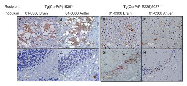
Figure 4. PrPSc (disease-associated form of prion protein)–specific immunohistochemistry in the brains of diseased mice. Transgenic (Tg) mice Tg(CerPrP)1536+/– inoculated with brain (A) and antler velvet (B) preparations from elk 01-0306 exhibit florid PrPSc-reactive plaques in the cerebral cortex at the level of the thalamus but retain integrity of cerebellar granular cells (C and D). Tg(CerPrP-E226)5037+/– mice inoculated with brain (E) and antler velvet (F) preparations from elk 01-0306 display small plaques and diffuse granular staining in the cerebral cortex, PrPSc deposition, and marked cerebellar neuronal loss (G and H).
Page created: December 16, 2010
Page updated: December 16, 2010
Page reviewed: December 16, 2010
The conclusions, findings, and opinions expressed by authors contributing to this journal do not necessarily reflect the official position of the U.S. Department of Health and Human Services, the Public Health Service, the Centers for Disease Control and Prevention, or the authors' affiliated institutions. Use of trade names is for identification only and does not imply endorsement by any of the groups named above.