Volume 8, Number 7—July 2002
Research
Emergence of Usutu virus, an African Mosquito-Borne Flavivirus of the Japanese Encephalitis Virus Group, Central Europe
Figure 1
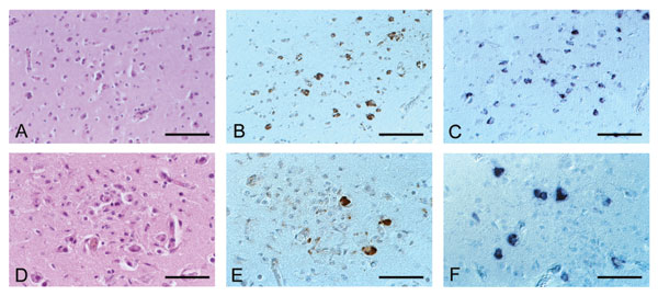
Figure 1. Histology and detection of viral signals in paraffin-embedded tissue sections of birds infected with Usutu virus (USUV). A–C, Eurasian Blackbird; D–F, Great Gray Owl; A, no histologic lesions present, hematoxylin and eosin staining; B immunohistochemistry, using a polyclonal antibody to West Nile virus, shows numerous positive neurons; C, in situ hybridization with USUV-specific oligonucleotide probe shows a staining pattern comparable with that in B; D, microglial nodule within the cerebral cortex, hematoxylin and eosin staining; E, immunohistochemistry shows single positive neurons within a glial nodule; F, in situ hybridization shows several positive neurons next to a glial nodule. Original magnification, x 130 (A–C), x 200 (D–F).