Volume 10, Number 11—November 2004
Research
Topographic Changes in SARS Coronavirus–infected Cells during Late Stages of Infection
Mah-Lee Ng* , J.W.M. Lee*, M.L.N. Leong*, A.-E. Ling†, H.-C. Tan‡, and E.E. Ooi‡
Figure 5
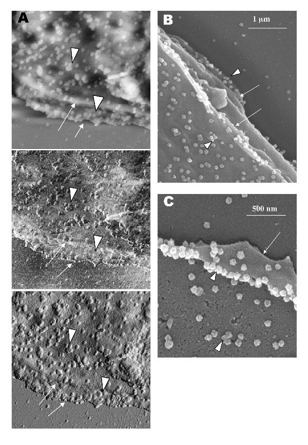
Figure 5. Vero cells infected with severe acute respiratory syndrome–associated coronavirus. A) An atomic force microscopy image of thickened, layered appearance of the edge of the cells (arrows), where active virus extrusion occurs. Arrowheads indicate the virus particles. B) The layered cell edge (arrows) seen by scanning electron microscopy. Virus particles extrude from the layered surfaces.
Page created: April 21, 2011
Page updated: April 21, 2011
Page reviewed: April 21, 2011
The conclusions, findings, and opinions expressed by authors contributing to this journal do not necessarily reflect the official position of the U.S. Department of Health and Human Services, the Public Health Service, the Centers for Disease Control and Prevention, or the authors' affiliated institutions. Use of trade names is for identification only and does not imply endorsement by any of the groups named above.