Volume 10, Number 3—March 2004
Research
Monkeypox Transmission and Pathogenesis in Prairie Dogs
Figure 2
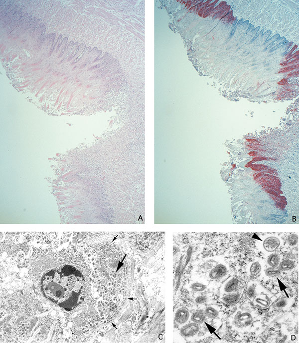
Figure 2. Ulcer on tongue of a prairie dog infected with monkeypox virus (A: hematoxylin and eosin stain, 12.5X original magnification). Orthopox viral antigens are abundant in the squamous epithelium, with lesser amounts in the ulcer bed (B: immunohistochemical stain using the anti-smallpox antibody, 12.5X original magnification). Tongue epithelial cell adjacent to epidermal basement membrane (small arrows) with Guarnieri-like inclusion (large arrow) (C: transmission electron microscopy, 2,400X original magnification). Higher magnification of the Guarnieri-like inclusion shows intracellular immature (arrowhead) and mature (arrows) orthopox virions. The mature virions consist of a dense core surrounded by several laminated zones and enclosed within an outer membrane. (D: transmission electron microscopy, 17,000X original magnification.)
1Veterinary Monkeypox Virus Working Group Members: CDC’s Infectious Diseases Pathology Activity: Patricia Greer, Michelle M. Packard, and William Lee, and CDC’s Poxvirus Section Laboratory: Victoria Olson, Richard Kline, Yu Li, and Linda Stempora.