Volume 10, Number 3—March 2004
Research
Monkeypox Transmission and Pathogenesis in Prairie Dogs
Figure 3
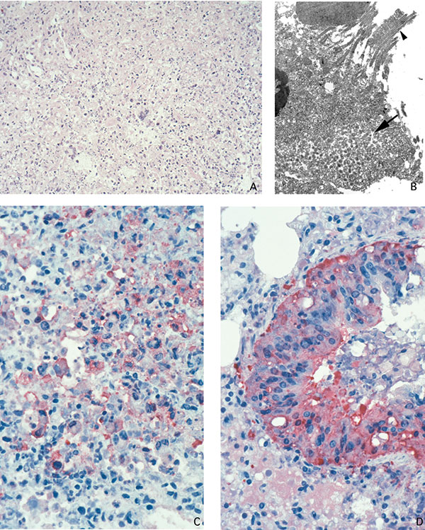
Figure 3. Lung of prairie dog infected with monkeypox virus, showing abundant intraalveolar mixed inflammatory infiltrate and necrosis (A: hematoxylin and eosin stain, 50X original magnification). Orthopox viral antigens are abundant in the cytoplasm of the bronchiolar epithelium (D: immunohistochemical assay anti–variola virus antibody, 100X original magnification). Macrophages, fibroblasts, and alveolar epithelial cells, as well as necrotic debris demonstrate orthopox viral antigens in pneumonic areas of the lung (C: immunohistochemical stain anti–variola virus antibody, 100X original magnification). Accumulation of intracellular mature virions (arrow) in bronchial epithelial cell (arrowhead pointing to cilia) (B: transmission electron microscopy, 2,400X original magnification).
1Veterinary Monkeypox Virus Working Group Members: CDC’s Infectious Diseases Pathology Activity: Patricia Greer, Michelle M. Packard, and William Lee, and CDC’s Poxvirus Section Laboratory: Victoria Olson, Richard Kline, Yu Li, and Linda Stempora.