Volume 10, Number 3—March 2004
Research
Monkeypox Transmission and Pathogenesis in Prairie Dogs
Jeannette Guarner* , Bill J. Johnson†, Christopher D. Paddock*, Wun-Ju Shieh*, Cynthia S. Goldsmith*, Mary G. Reynolds*, Inger K. Damon*, Russell L. Regnery*, Sherif R. Zaki*, and the Veterinary Monkeypox Virus Working Group
Figure 1
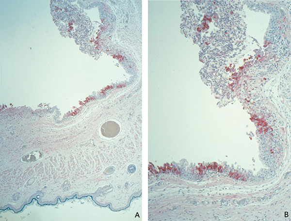
Figure 1. Immunohistochemical staining of a prairie dog eyelid infected with monkeypox virus, showing orthopox virus antigen staining of the cytoplasm of the epithelium of the palpebral conjunctivae (assay using anti–variola virus antibody; original magnifications: A, 12.5X; B, 25X).
1Veterinary Monkeypox Virus Working Group Members: CDC’s Infectious Diseases Pathology Activity: Patricia Greer, Michelle M. Packard, and William Lee, and CDC’s Poxvirus Section Laboratory: Victoria Olson, Richard Kline, Yu Li, and Linda Stempora.
Page created: February 08, 2011
Page updated: February 08, 2011
Page reviewed: February 08, 2011
The conclusions, findings, and opinions expressed by authors contributing to this journal do not necessarily reflect the official position of the U.S. Department of Health and Human Services, the Public Health Service, the Centers for Disease Control and Prevention, or the authors' affiliated institutions. Use of trade names is for identification only and does not imply endorsement by any of the groups named above.