Volume 11, Number 10—October 2005
Research
Pyrosequencing Bacillus anthracis
Cite This Article
Citation for Media
Abstract
Pyrosequencing technology is a sequencing method that screens DNA nucleotide incorporation in real time. A set of coupled enzymatic reactions, together with bioluminescence, detects incorporated nucleotides in the form of light pulses, which produces a profile of characteristic peaks in a pyrogram. We used this technology to identify the warfare agent Bacillus anthracis by sequencing 4 single nucleotide polymorphisms (SNPs) in the rpoB gene as chromosomal markers for B. anthracis. In addition, 1 segment in each of the B. anthracis plasmids pXO1 and pXO2 was analyzed to determine the virulence status of the bacterial strains. Pyrosequencing technology is a powerful method to identify B. anthracis.
Bacillus anthracis is a gram-positive, rod-shaped, spore-forming bacterium that causes the acute mammalian disease anthrax when endospores enter the body. The infection is often fatal if not treated with antimicrobial drugs before or when the first symptoms appear. The bacterium can infect livestock and humans by gastrointestinal, cutaneous, or respiratory routes. Potentially, B. anthracis spores can be an effective biological weapon because of their high stability. They do not divide, have no metabolism, and are resistant to drying, heat, UV light, and many disinfectants. In 2001, letters containing anthrax spores caused illness in 22 persons, leading to 5 deaths in the United States.
B. anthracis has 2 plasmids: the toxin-encoding pXO1 (182 kb) and capsule-encoding pXO2 (95 kb) (1,2). Both plasmids are required for virulence; lacking 1 of the plasmids attenuates the microorganism. The pXO1 plasmid contains genes lef, cya, and pag, which encode the toxin's lethal factor, edema factor, and protective antigen, respectively (3–5). The pXO2 plasmid contains the genes capA, capB, and capC, necessary for capsule formation (6). These genes have been used as markers to identify B. anthracis with polymerase chain reaction (PCR) in both environmental and clinical samples (7–9).
Differentiating between B. anthracis and closely related B. cereus and B. thuringiensis is difficult (10). Usually, phenotypic characteristics, such as susceptibility to β-lactam antimicrobial drugs, lack of hemolysis, lack of motility on sheep blood agar plate, and inability to ferment salicin, are used to differentiate (11,12). A variety of chromosomal markers that appear to be specific have been suggested for genotypic species determination of B. anthracis (13–18). We studied single nucleotide polymorphisms (SNPs) in the rpoB gene, described by Qi et al. (13) by using pyrosequencing technology (19). This technology can determine SNPs and short DNA stretches in real time, starting from PCR products. Biotinylated PCR amplicons that cover the region of interest are immobilized onto solid streptavidin coated beads and converted to single-stranded form. A sequencing primer is hybridized to the single-stranded DNA, and incorporation of added nucleotides is detected as light peaks by an enzymatic cascade. Enzymatic degradation of excess nucleotides allows the reaction to be performed in a single tube. When one starts from PCR products, <96 genetic targets can be sequenced within 1 hour.
In this study, we used the rpoB gene as a chromosomal marker to discriminate between B. anthracis and closely related bacillus species. We studied 4 B. anthracis–specific rpoB SNPs located at positions 911, 912, 913, and 914 in duplex sequencing reactions by using a unique sequencing primer for each desired SNP in a collection of 17 anthracis and 10 non-anthracis Bacillus strains. Simultaneously, we investigated the distribution of virulence plasmids pXO1 and pXO2 among these strains by using PCR and pyrosequencing technology to rapidly verify the amplicons.
The B. anthracis reference strains used in this study were obtained from the National Collection of Type Cultures, London, England, and the Swedish Defense Research Agency. Reference strains of B. cereus, B. mycoides, and B. thuringiensis were obtained from the Culture Collection University of Gothenburg of Sweden. All bacterial strains are listed in Table 1. Bacteria were cultured on blood sheep agar at 37°C for 16 h, and genomic DNA was prepared by using a commercially available DNA extraction kit, QIAamp tissue protocol (Hilden, Stockholm, Sweden). The DNA was boiled at 99°C for 15 min, plated on blood agar, and incubated for 3 days. No growth was observed, and the DNA was removed from the biosafety level 3 laboratory. All material, including the DNA, is under the protection of our institute.
PCR
All reagents used for amplification of bacterial DNA were from Amersham Biosciences (Uppsala, Sweden) except for primers, which were from Invitrogen Life Technologies (Paisley, United Kingdom). Table 2 shows the primer sequences. The reverse primer for each PCR fragment was biotinylated. PCR primers were designed to amplify a 176-bp fragment of rpoB, 179 bp of the pXO1 plasmid, and 127 bp of the pXO2 plasmid. PCR was performed in 50-μL reaction mixtures containing 1× PCR buffer (10 mmol/L Tris-HCl, pH 8.3, 50 mmol/L KCl, 2.5 mmol/L MgCl2), 0.8 U Taq DNA polymerase, 0.2 mmol/L each nucleotide, 0.1 μmol/L each primer and 5 μL eluate containing DNA. The reaction mixture was subjected to 95°C for 5 min and 45 cycles of 95°C for 30 s, annealing at 60°C for 30 s, and elongation at 72°C for 30 s, followed by terminal extension at 72°C for 7 min.
Pyrosequencing Analysis
Two primers were designed to sequence of 30 nucleotides within the PCR amplicons generated from plasmids pXO1 and pXO2, respectively. In addition, 1 sequencing primer was designed for each of the 4 rpoB SNPs to be determined (Table 2). The rpoB primers were used in duplex sequencing reactions so that primers 911 and 912 were combined in a single reaction for sequencing of SNPs at positions 911 and 912, while primers 913 and 914 were used for combined sequencing of SNPs at positions 913 and 914. For sequencing according to the pyrosequencing technology, biotinylated PCR amplicons were immobilized onto streptavidin-coated magnetic beads and denatured to produce single-stranded DNA by using a PSQ 96 Sample Prep Tool (Biotage AB, Uppsala, Sweden). Sequencing primers were added and allowed to hybridize to the strands, after which sequencing was performed according to the manufacturer's instructions. All steps were performed at room temperature.
Bacterial Strains and DNA Extraction
Figure
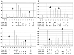
Figure. Sequence analysis of single nucleotide polymorphisms (SNPs) in the rpoB gene of Bacillus anthracis National Collection of Type Cultures (NCTC) 2026 (A and B) and B. cereus Culture Collection University of...
B. anthracis was unambiguously identified by determining 4 SNPs in the rpoB gene by using pyrosequencing technology; thus, we were able to distinguish B. anthracis from the other tested bacillus species. The Figure shows representative output diagrams, or pyrograms, from duplex sequencing reactions of the SNPs in B. anthracis National Collection of Type Cultures 2026 and B. cereus Culture Collection University of Gothenburg 7414. The specific nucleotides 911C, 912T, 913C, and 914A were found in all B. anthracis strains tested and appear to be unique to B. anthracis. The corresponding nucleotides in all tested non-anthracis strains (B. cereus, B. thuringiensis, and B. mycoides) were 911T, 912C, 913T, and 914G. In duplex sequencing reactions, we could easily determine 2 SNPs in each pyrogram (Figure).Presence of the virulence plasmids was determined by PCR and sequence verification of regions on plasmids pXO1 and pXO2 (Table 1). The assay was applied on 17 B. anthracis strains, 7 B. cereus strains, 2 B. thuringiensis strains, and 1 B. mycoides strain. Of the 17 B. anthracis isolates investigated, 9 isolates were positive for both pXO1 and pXO2, while the remaining 8 lacked either 1 or both of the virulence plasmids. All non-anthracis strains were negative for both plasmids. pXO1 and pXO2 PCR amplicons originating from the B. anthracis strains were verified by sequencing 30 nucleotides with the pyrosequencing technology. The nucleotide sequence following the sequencing primers located within the amplified fragments was AAGATATTATCAAGGGATATTTTAAGTAAA for all pXO1 amplicons and ACCACTCATTAAGTTCTTCGCACCGCTAAA for all pXO2 amplicons, which agreed with all nucleotide sequences of these regions submitted to the GenBank nucleotide database (http://www.ncbi.nlm.nih.gov), accession numbers AF065404, AE011190, AJ413934, AE017336, AJ413935, M29081, M30210, AF188935, AE011191, and AE017335. B. anthracis was successfully identified by using pyrosequencing technology for genotyping 4 SNP positions of the rpoB gene, which appear to be specific for B. anthracis, and 2 fragments of virulence plasmids pXO1 and pXO2.
In biologic warfare, speed and accuracy are in high demand for identifying and characterizing microbial species. In this study, we investigated the possibility of using pyrosequencing technology to rapidly identify and characterize strains of B. anthracis and distinguish them from related non-anthracis Bacillus strains. This method has been used to analyze multiple targets that are important in microbial infections (20,21).
By determining 4 SNPs in the rpoB gene, B. anthracis strains were successfully identified. This chromosomal marker can discriminate between B. anthracis and other closely related species from the Bacillus genus. The 16S rRNA gene cannot be relied upon to differentiate B. anthracis from its close relatives; therefore, we did not include this target in the assay (22). By careful design of nucleotide dispensation order, multiple SNPs may be analyzed in 1 single sequencing reaction by using a unique primer for each desired SNP. To save time and reduce reagent cost, we analyzed the rpoB SNPs in duplex pyrosequencing reactions. The resulting pyrograms of overlapping sequences were easily resolved by the accompanying software (Figure). Using 1 well for all 4 SNP positions may further optimize the method.
This technology validates PCR-based assays by qualitatively verifying that a positive PCR result is not the effect of nonspecific amplification, as shown here by sequence verification of PCR amplicon generated from virulence plasmids pXO1 and pXO2. The risk of false-positive results is thereby minimized.
We illustrate for the first time how pyrosequencing technology can identify B. anthracis. Using this technology in diagnostic laboratories is advantageous because it is rapid, simple, nonradioactive, inexpensive, and automated. It is a powerful method to rapidly determine genetic targets; as many as 96 samples can be analyzed in 40 minutes. Genetic analysis with pyrosequencing technology could make selecting antimicrobial drug treatment easier and potentially complement typing methods and time-consuming, traditional microbial identification, such as biochemical testing, phage lysing assays, and immunologic assays.
Ms Wahab is a microbiologist at the Centre for Microbiological Preparedness at the Swedish Institute for Infectious Disease Control. She works mainly with diagnostics and developing new techniques to identify bacterial biosafety level 3 organisms.
Acknowledgments
We thank Hong-Yan Zhang for providing the resources to perform this study and the Swedish Defense Research Agency for providing Bacillus strains.
This work was supported by grants from the Swedish Emergency Management Agency.
References
- Mikesell P, Ivins BE, Ristroph JD, Dreier TM. Evidence for plasmid-mediated toxin production in Bacillus anthracis. Infect Immun. 1983;39:371–6.PubMedGoogle Scholar
- Uchida I, Sekizaki T, Hashimoto K, Terakado N. Association of the encapsulation of Bacillus anthracis with a 60 megadalton plasmid. J Gen Microbiol. 1985;131:363–7.PubMedGoogle Scholar
- Bragg TS, Robertson DL. Nucleotide sequence and analysis of the lethal factor gene (lef) from Bacillus anthracis. Gene. 1989;81:45–54. DOIPubMedGoogle Scholar
- Robertson DL, Tippetts MT, Leppla SH. Nucleotide sequence of the Bacillus anthracis edema factor gene (cya): a calmodulin-dependent adenylate cyclase. Gene. 1988;73:363–71. DOIPubMedGoogle Scholar
- Welkos SL, Lowe JR, Eden-McCutchan F, Vodkin M, Leppla SH, Schmidt JJ. Sequence and analysis of the DNA encoding protective antigen of Bacillus anthracis. Gene. 1988;69:287–300. DOIPubMedGoogle Scholar
- Makino SI, Uchida I, Terakado N, Sasakawa C, Yoshikakawa M. Molecular characterization and protein analysis of the cap region, which is essential for encapsulation in Bacillus anthracis. J Bacteriol. 1989;171:722–30.PubMedGoogle Scholar
- Makino SI, Iinuma-Okada Y, Maruyama T, Ezaki T, Sasakawa C, Yoshikawa M. Direct detection of Bacillus anthracis DNA in animals by polymerase chain reaction. J Clin Microbiol. 1993;31:547–51.PubMedGoogle Scholar
- Ramisse V, Patra G, Garrigue H, Guesdon JL, Mock M. Identification and characterization of Bacillus anthracis by multiplex PCR analysis of sequences on plasmids pXO1 and pXO2 and chromosomal DNA. FEMS Microbiol Lett. 1996;145:9–16. DOIPubMedGoogle Scholar
- Sjöstedt A, Eriksson U, Ramisse V, Garrigue H. Detection of Bacillus anthracis spores in soil by PCR. FEMS Microbiol Ecol. 1997;23:159–68. DOIGoogle Scholar
- Ash C, Farrow JAE, Dorsch M, Stackebrandt E, Collins MD. Comparative analysis of Bacillus anthracis, Bacillus cereus, and related species on the basis of reverse transcriptase sequencing of 16S rRNA. Int J Syst Bacteriol. 1991;41:343–6. DOIPubMedGoogle Scholar
- Logan NA, Carman J, Melling J, Berkeley R. Identification of Bacillus anthracis by API tests. J Med Microbiol. 1985;20:75–85. DOIPubMedGoogle Scholar
- Dixon TC, Meselson M, Guilleman J, Hanna P. Anthrax. N Engl J Med. 1999;341:815–26. DOIPubMedGoogle Scholar
- Qi Y, Patra G, Liang X, Williams LE, Rose S, Redkar RJ, Utilization of the rpoB gene as a specific chromosomal marker for real-time PCR detection of Bacillus anthracis. Appl Environ Microbiol. 2001;67:3720–7. DOIPubMedGoogle Scholar
- Ellerbrock H, Nattermann H, Özel M, Beutin L, Appel B, Pauli G. Rapid and sensitive identification of pathogenic and apathogenic Bacillus anthracis by real-time PCR. FEMS Microbiol Lett. 2002;214:51–9. DOIPubMedGoogle Scholar
- Van Ert MN, Hofstadler SA, Jiang Y, Busch JD, Wagner DM, Drader JJ, Mass spectrometry provides accurate characterization of two genetic marker types in Bacillus anthracis. Biotechniques. 2004;37:642–4.PubMedGoogle Scholar
- Hurtle W, Bode E, Kulesh DA, Kaplan RS, Garrison J, Bridge D, Detection of the Bacillus anthracis gyrA gene by using a minor groove binder probe. J Clin Microbiol. 2004;42:179–85. DOIPubMedGoogle Scholar
- Pearson T, Busch JD, Ravel J, Read TD, Rhoton SD, U'Ren JM, Phylogenetic discovery bias in Bacillus anthracis using single-nucleotide polymorphisms from whole-genome sequencing. Proc Natl Acad Sci U S A. 2004;101:13536–41. DOIPubMedGoogle Scholar
- Hill KK, Ticknor LO, Okinaka RT, Asay M, Blair H, Bliss KA, Fluorescent amplified fragment length polymorphism analysis of Bacillus anthracis, B. cereus, and B. thuringiensis isolates. Appl Environ Microbiol. 2004;70:1068–80. DOIPubMedGoogle Scholar
- Ronaghi M, Uhlen M, Nyren P. A sequencing method based on real time pyrophosphate. Science. 1998;281:363, 365.
- Hjalmarsson S, Alderborn A, Fock C, Muldin I, Kling H, Uhlen M, Rapid combined characterization of microorganism and host genotypes using a single technology. Helicobacter. 2004;9:138–45. DOIPubMedGoogle Scholar
- Sinclair A, Arnold C, Woodford N. Rapid detection and estimation by pyrosequencing of 23S rRNA genes with a single nucleotide polymorphism conferring linezolid resistance in enterococci. Antimicrob Agents Chemother. 2003;47:3620–2. DOIPubMedGoogle Scholar
- Blackwood KS, Turenne CY, Harmsen D, Kabani AM. Reassessment of sequence-based targets for identification of Bacillus species. J Clin Microbiol. 2004;42:1626–30. DOIPubMedGoogle Scholar
Figure
Tables
Cite This ArticleTable of Contents – Volume 11, Number 10—October 2005
| EID Search Options |
|---|
|
|
|
|
|
|
Please use the form below to submit correspondence to the authors or contact them at the following address:
Lars Engstrand, Section of Bacteriology, Swedish Institute for Infectious Disease Control, SE 171 82, Solna, Sweden; fax: 46-8-30-17-97
Top