Skip directly to site content Skip directly to page options Skip directly to A-Z link Skip directly to A-Z link Skip directly to A-Z link
Volume 11, Number 4—April 2005
Research

Experimental Infection of Prairie Dogs with Monkeypox Virus

Shu-Yuan Xiao*, Elena Sbrana*, Douglas M. Watts*, Marina Siirin*, Amelia P.A. Travassos da Rosa*, and Robert B. Tesh*Comments to Author 
Author affiliations: *University of Texas Medical Branch, Galveston, Texas, USA

Main Article

Figure 2

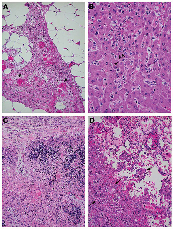
Photomicrographs of pathologic changes in tissues of prairie dogs infected with monkeypox virus. A) Abdominal adipose tissue from an intraperitoneally infected animal showing focal vasculitis (arrowheads), necrosis, and proliferation of fibroblasts. B) Mild hepatitis, characterized by focal inflammatory cell infiltration in the lobules and hepatocytes containing cytoplasmic inclusion bodies (arrowheads). C) Severe necrosis of the thymus from an animal infected intranasally. The necrotic areas co

Figure 2. Photomicrographs of pathologic changes in tissues of prairie dogs infected with monkeypox virus. A) Abdominal adipose tissue from an intraperitoneally infected animal showing focal vasculitis (arrowheads), necrosis, and proliferation of fibroblasts. B) Mild hepatitis, characterized by focal inflammatory cell infiltration in the lobules and hepatocytes containing cytoplasmic inclusion bodies (arrowheads). C) Severe necrosis of the thymus from an animal infected intranasally. The necrotic areas contain many swollen macrophages with cytoplasm full of intensively eosinophilic material (viral proteins). D) Lung from the same animal, showing inflammation with swollen cells (arrowheads), alveolar edema, and necrosis of the pleura. The latter is infiltrated by many inclusion-filled macrophages and proliferating fibroblasts (the thick layer between the 2 arrows). Magnification: A, 10× objective; B, 40× objective; C, 10× objective; D, 20× objective. Hematoxylin and eosin stain.

Main Article

Page created: May 23, 2011
Page updated: May 23, 2011
Page reviewed: May 23, 2011
The conclusions, findings, and opinions expressed by authors contributing to this journal do not necessarily reflect the official position of the U.S. Department of Health and Human Services, the Public Health Service, the Centers for Disease Control and Prevention, or the authors' affiliated institutions. Use of trade names is for identification only and does not imply endorsement by any of the groups named above.
file_external