Volume 11, Number 4—April 2005
Research
Experimental Infection of Prairie Dogs with Monkeypox Virus
Shu-Yuan Xiao*, Elena Sbrana*, Douglas M. Watts*, Marina Siirin*, Amelia P.A. Travassos da Rosa*, and Robert B. Tesh*
Figure 3
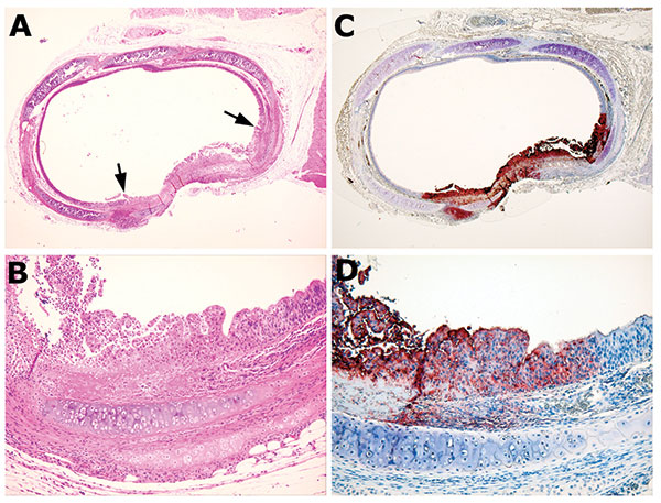
Figure 3. Bronchus from animal MPX-9, which was infected intranasally. A) Cross-section of a bronchus, showing focal metaplasia and proliferation (between the arrows) of the luminal epithelium. B) Higher magnification showing the details of the metaplastic epithelium, accompanied by focal necrosis. Compare to the adjacent unaffected area, which is lined by normal ciliated columnar epithelial cells. C and D) Immunohistochemical staining of the corresponding field shows presence of viral antigen limited to the region of epithelial abnormality. A and B, hematoxylin and eosin stain; C and D, immunoperoxidase staining with vaccinia antibody. Original magnification: A and C, 4× objectives; B and D, 20× objectives.
Page created: May 23, 2011
Page updated: May 23, 2011
Page reviewed: May 23, 2011
The conclusions, findings, and opinions expressed by authors contributing to this journal do not necessarily reflect the official position of the U.S. Department of Health and Human Services, the Public Health Service, the Centers for Disease Control and Prevention, or the authors' affiliated institutions. Use of trade names is for identification only and does not imply endorsement by any of the groups named above.