Volume 14, Number 3—March 2008
Research
Discovering and Differentiating New and Emerging Clonal Populations of Chlamydia trachomatis with a Novel Shotgun Cell Culture Harvest Assay
Naraporn Somboonna*†, Sally Mead*, Jessica Liu†, and Deborah Dean*†‡
Figure 1
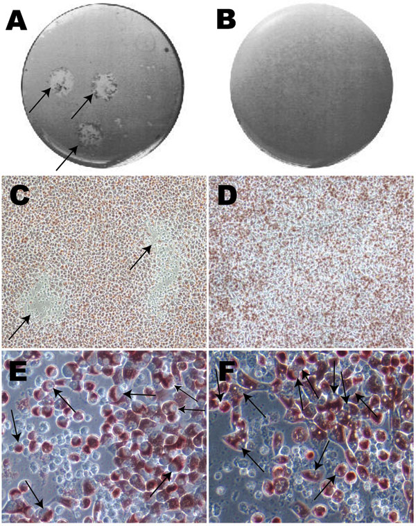
Figure 1. Photographs and optical microscopy views of the wells showing plaques formed by Chlamydia trachomatis F/IC-Cal3 (A, C, E) and no plaque formed by clinical F persistent strain (B, D, F). A) Single well showing 2 distinct plaques (indicated by arrows). B) Well showing no plaque morphology. C) and D) Optical microscopy image showing plaque areas with little or no neutral red staining (arrows) surrounded by viable cells stained red (magnification ×100). Higher magnification (×400) showed numerous cells that had been infected by reference strain F/IC-Cal3 (E) and the clinical persistent F strain (F).
Page created: July 07, 2010
Page updated: July 07, 2010
Page reviewed: July 07, 2010
The conclusions, findings, and opinions expressed by authors contributing to this journal do not necessarily reflect the official position of the U.S. Department of Health and Human Services, the Public Health Service, the Centers for Disease Control and Prevention, or the authors' affiliated institutions. Use of trade names is for identification only and does not imply endorsement by any of the groups named above.