Skip directly to site content Skip directly to page options Skip directly to A-Z link Skip directly to A-Z link Skip directly to A-Z link
Volume 14, Number 3—March 2008
Research

Discovering and Differentiating New and Emerging Clonal Populations of Chlamydia trachomatis with a Novel Shotgun Cell Culture Harvest Assay

Naraporn Somboonna*†, Sally Mead*, Jessica Liu†, and Deborah Dean*†‡Comments to Author 
Author affiliations: *Children’s Hospital Oakland Research Institute, Oakland, California, USA; †University of California, Berkeley, California, USA; ‡University of California School of Medicine, San Francisco, California, USA;

Main Article

Figure 3

Photographs of light and fluorescent microscopy showing the shotgun cell culture harvest method for isolating clonal populations of clinically persistant strains. A) 1 well in a 6-well plate after harvesting 10 random areas using sterile pipettes; no plaques could be visualized in any well unlike the situation for the reference strains in Figure 2. B) The same well before harvesting infected areas (magnification ×100). C) Light microscopy view of an area with infected cells containing small inclusion bodies after agarose overlays had been removed (magnification ×400). D) Fluorescent microscopy view of the same field as in C (magnification ×400); infected cells were stained with Chlamydia trachomatis–specific lipopolysaccharide antibodies. Arrows denote small fluorescing inclusion bodies within the cell cytoplasm.

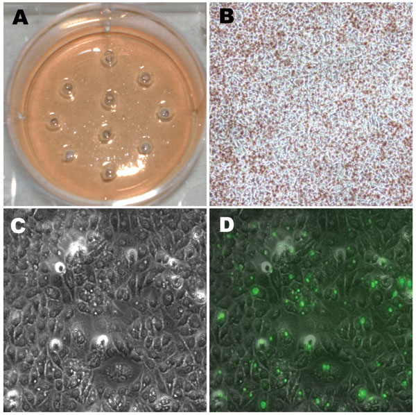
Figure 3. Photographs of light and fluorescent microscopy showing the shotgun cell culture harvest method for isolating clonal populations of clinically persistant strains. A) 1 well in a 6-well plate after harvesting 10 random areas using sterile pipettes; no plaques could be visualized in any well unlike the situation for the reference strains in Figure 2. B) The same well before harvesting infected areas (magnification ×100). C) Light microscopy view of an area with infected cells containing small inclusion bodies after agarose overlays had been removed (magnification ×400). D) Fluorescent microscopy view of the same field as in C (magnification ×400); infected cells were stained with Chlamydia trachomatis–specific lipopolysaccharide antibodies. Arrows denote small fluorescing inclusion bodies within the cell cytoplasm.

Main Article

Page created: July 07, 2010
Page updated: July 07, 2010
Page reviewed: July 07, 2010
The conclusions, findings, and opinions expressed by authors contributing to this journal do not necessarily reflect the official position of the U.S. Department of Health and Human Services, the Public Health Service, the Centers for Disease Control and Prevention, or the authors' affiliated institutions. Use of trade names is for identification only and does not imply endorsement by any of the groups named above.
file_external