Volume 14, Number 4—April 2008
Dispatch
Neuroinvasion by Mycoplasma pneumoniae in Acute Disseminated Encephalomyelitis
Bernhard Stamm* , Michael Moschopulos*, Hansjoerg Hungerbuehler*, Jeannette Guarner†, Gillian L. Genrich†, and Sherif R. Zaki†
Figure
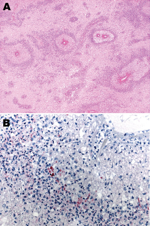
Figure. A) Subcortical cerebral white matter with numerous perivascular foci of demyelination and necrosis (hematoxylin and eosin stain, original magnification ×40). B) Immunohistochemical evidence of Mycoplasma pneumoniae antigen inside macrophages present in the perivascular inflammatory infiltrate (immunohistochemical assay performed by using the monoclonal anti–M. pneumoniae antibody and naphthol fast red as counterstain, original magnification ×100).
Page created: July 09, 2010
Page updated: July 09, 2010
Page reviewed: July 09, 2010
The conclusions, findings, and opinions expressed by authors contributing to this journal do not necessarily reflect the official position of the U.S. Department of Health and Human Services, the Public Health Service, the Centers for Disease Control and Prevention, or the authors' affiliated institutions. Use of trade names is for identification only and does not imply endorsement by any of the groups named above.