Volume 14, Number 4—April 2008
Research
Wild Ducks as Long-Distance Vectors of Highly Pathogenic Avian Influenza Virus (H5N1)
Appendix Figure 4
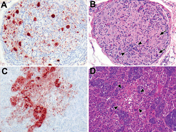
Appendix Figure 4. Highly pathogenic avian influenza virus (H5N1) infection in tufted ducks. A) Neurons and satellite cells in a mesenteric ganglion expressing abundant influenza virus antigen. B) Ganglioneuritis in the same mesenteric ganglion, characterized by neuronal necrosis (arrows) and lymphocyte infiltration (between arrowheads). C) Expression of influenza virus antigen in medullary and cortical cells of an adrenal gland and D) focal necrosis, characterized by hypereosinophilia, pyknosis, and vacuolization (between arrowheads). Original magnification ×100. Tissues were stained either by immunohistochemistry that used a monoclonal antibody against the nucleoprotein of influenza A virus as a primary antibody (A, C) or with hematoxylin and eosin (B, D).