Skip directly to site content Skip directly to page options Skip directly to A-Z link Skip directly to A-Z link Skip directly to A-Z link
Volume 14, Number 4—April 2008
Research

Wild Ducks as Long-Distance Vectors of Highly Pathogenic Avian Influenza Virus (H5N1)

Juthatip Keawcharoen*, Debby van Riel*, Geert van Amerongen*, Theo M. Bestebroer*, Walter E. Beyer*, Rob van Lavieren*, Albert D.M.E. Osterhaus*, Ron A.M. Fouchier*, and Thijs Kuiken*Comments to Author 
Author affiliations: *Erasmus Medical Center, Rotterdam, the Netherlands;

Main Article

Figure 3

Mean cloacal excretion of highly pathogenic avian influenza virus (H5N1) by wild ducks by A) virus isolation and C) reverse transcription–PCR (RT-PCR). Legend for panels A–D as in Figure 2. E) Pancreas showing multiple foci of necrosis (between arrowheads) in a pochard. F) Pancreatic acinar cells in a pochard and H) hepatocytes in a tufted duck, showing the transition area between normal and necrotic tissue expressing abundant influenza virus antigen. G) Pancreatic lesions in a pochard and I) hepatic lesions in a tufted duck, characterized by sharp transition between normal tissue (left side of panels) and foci of necrosis and inflammatory cell infiltration (right side of panels). F, G original magnification ×50. H, I original magnification ×100. Tissues were stained either by immunohistochemistry that used a monoclonal antibody against the nucleoprotein of influenza A virus as a primary antibody (F, H) or with hematoxylin and eosin (G, I).

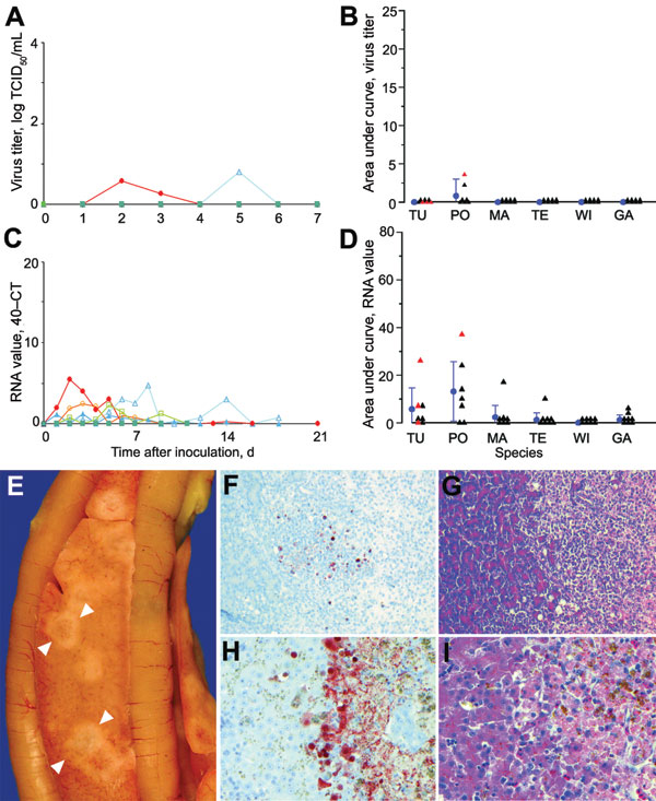
Figure 3. Mean cloacal excretion of highly pathogenic avian influenza virus (H5N1) by wild ducks by A) virus isolation and C) reverse transcription–PCR (RT-PCR). Legend for panels A–D as in Figure 2. E) Pancreas showing multiple foci of necrosis (between arrowheads) in a pochard. F) Pancreatic acinar cells in a pochard and H) hepatocytes in a tufted duck, showing the transition area between normal and necrotic tissue expressing abundant influenza virus antigen. G) Pancreatic lesions in a pochard and I) hepatic lesions in a tufted duck, characterized by sharp transition between normal tissue (left side of panels) and foci of necrosis and inflammatory cell infiltration (right side of panels). F, G original magnification ×50. H, I original magnification ×100. Tissues were stained either by immunohistochemistry that used a monoclonal antibody against the nucleoprotein of influenza A virus as a primary antibody (F, H) or with hematoxylin and eosin (G, I).

Main Article

Page created: July 13, 2010
Page updated: July 13, 2010
Page reviewed: July 13, 2010
The conclusions, findings, and opinions expressed by authors contributing to this journal do not necessarily reflect the official position of the U.S. Department of Health and Human Services, the Public Health Service, the Centers for Disease Control and Prevention, or the authors' affiliated institutions. Use of trade names is for identification only and does not imply endorsement by any of the groups named above.
file_external