Skip directly to site content Skip directly to page options Skip directly to A-Z link Skip directly to A-Z link Skip directly to A-Z link
Volume 14, Number 4—April 2008
Research

Retrospective Analysis of Monkeypox Infection

Melissa E. Dubois* and Mark K. Slifka*Comments to Author 
Author affiliations: *Oregon Health and Science University, Beaverton, Oregon, USA;

Main Article

Figure 2

Development of a monkeypox (MPV)–specific diagnostic assay using Western blot analysis. Adsorption of cross-reactive orthopoxvirus antibodies with vaccinia antigen before Western blot analysis provided easier identification of monkeypox-specific bands. A) Two micrograms of sucrose gradient–purified monkeypox virus or vaccinia virus (VV) were separated by sodium dodecyl sulfate–polyacrylamide gel electrophoresis (4%–20% gels) and stained with GelCode Blue (Pierce, Rockford, IL, USA) to compare banding patterns and confirm equivalent protein loading. Proteins were electrophoretically transferred to polyvinylidene difluoride membranes and probed with plasma from B) a monkeypox-immune person, C) a vaccinia-immune person, or D) an orthopoxvirus-naive person after adsorption of plasma with control antigen (uninfected H2O2-treated BSC40 cell lysate) or vaccinia antigen (H2O2-inactived vaccinia-infected BSC40 cell lysate). Immunoreactive bands were detected with peroxidase-conjugated antihuman immunoglobulin G plus chemiluminescent substrate and exposed to x-ray film. Arrows indicate location of diagnostic bands with apparent molecular masses of 148, 124, and 39 kDa. Rectangles indicate locations of diagnostic bands.

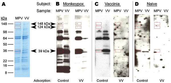
Figure 2. Development of a monkeypox (MPV)–specific diagnostic assay using Western blot analysis. Adsorption of cross-reactive orthopoxvirus antibodies with vaccinia antigen before Western blot analysis provided easier identification of monkeypox-specific bands. A) Two micrograms of sucrose gradient–purified monkeypox virus or vaccinia virus (VV) were separated by sodium dodecyl sulfate–polyacrylamide gel electrophoresis (4%–20% gels) and stained with GelCode Blue (Pierce, Rockford, IL, USA) to compare banding patterns and confirm equivalent protein loading. Proteins were electrophoretically transferred to polyvinylidene difluoride membranes and probed with plasma from B) a monkeypox-immune person, C) a vaccinia-immune person, or D) an orthopoxvirus-naive person after adsorption of plasma with control antigen (uninfected H2O2-treated BSC40 cell lysate) or vaccinia antigen (H2O2-inactived vaccinia-infected BSC40 cell lysate). Immunoreactive bands were detected with peroxidase-conjugated antihuman immunoglobulin G plus chemiluminescent substrate and exposed to x-ray film. Arrows indicate location of diagnostic bands with apparent molecular masses of 148, 124, and 39 kDa. Rectangles indicate locations of diagnostic bands.

Main Article

Page created: July 13, 2010
Page updated: July 13, 2010
Page reviewed: July 13, 2010
The conclusions, findings, and opinions expressed by authors contributing to this journal do not necessarily reflect the official position of the U.S. Department of Health and Human Services, the Public Health Service, the Centers for Disease Control and Prevention, or the authors' affiliated institutions. Use of trade names is for identification only and does not imply endorsement by any of the groups named above.
file_external