Volume 14, Number 4—April 2008
Research
Retrospective Analysis of Monkeypox Infection
Figure 3
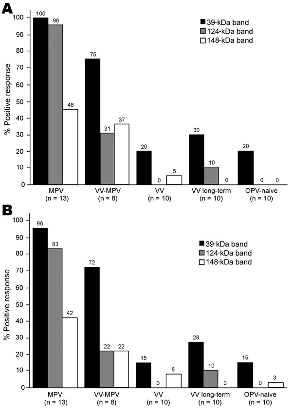
Figure 3. Diagnosis of monkeypox infection by Western blot analysis. Plasma samples from unvaccinated monkeypox-infected (2–30 months postinfection) (MPV), vaccinia-immune monkeypox-infected (2–6 months postinfection) (VV-MPV), primary vaccinia-immune (2–4 months postimmunization) (VV), long-term vaccinia-immune (>20 years postimmunization) (VV long-term), and orthopoxvirus-naive (OPV) persons were analyzed by Western blot after adsorption with vaccinia-infected BSC40 cell lysate to reduce cross-reactive antibodies as described in Figure 2. Immunoreactivity to diagnostic protein bands of ≈39 kDa, 124 kDa, and 148 kDa was assessed by A) unblinded analysts with knowledge of subject medical history (n = 2) and B) blinded analysts who did not have knowledge of subject medical history (n = 4). Findings of each analyst were averaged for each person and percentages shown represent a composite of all data points.