Volume 14, Number 4—April 2008
Dispatch
Human Thelaziasis, Europe
Domenico Otranto* and Moreno Dutto†
Figure 2
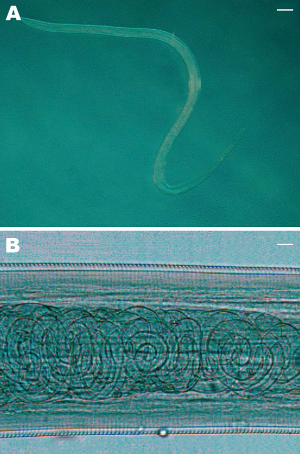
Figure 2. A) Female Thelazia callipaeda isolated from patient 4. The posterior end is on the left and the anterior end is on the right (magnification × 200). Scale bar = 500 μm. B) T. callipaeda mature first-stage larvae in the distal uterus (magnification ×100). Scale bar = 30 μm.
Page created: July 14, 2010
Page updated: July 14, 2010
Page reviewed: July 14, 2010
The conclusions, findings, and opinions expressed by authors contributing to this journal do not necessarily reflect the official position of the U.S. Department of Health and Human Services, the Public Health Service, the Centers for Disease Control and Prevention, or the authors' affiliated institutions. Use of trade names is for identification only and does not imply endorsement by any of the groups named above.