Volume 14, Number 6—June 2008
Letter
Bovine Kobuviruses from Cattle with Diarrhea
Cite This Article
Citation for Media
To the Editor: A new species of kobuvirus, named U-1 strain, was first recognized in 2003 as a cytopathic contaminant in a culture medium of HeLa cells that had been used for >30 years in the laboratory (1). The RNA genome of the U-1 strain comprises 8,374 nucleotides; the genome organization is analogous to that of picornaviruses. Morphologically, the U-1 strain resembles the Aichi virus, but genetically it is distinct (1). Therefore, the U-1 strain is classified as a new species of genus Kobuvirus in the family Picornaviridae, and it is called bovine kobuvirus (1). To date, the genus Kobuvirus consists of 2 species, Aichi virus and bovine kobuvirus (2). The Aichi virus is associated with acute gastroenteritis in humans (3–5); bovine kobuvirus infection has been detected only in cattle (1).
Only 1 report has described the discovery and epidemiologic features of bovine kobuvirus (1). Of serum samples from 72 healthy cattle, 43 (59.7%) were positive for neutralizing antibody against bovine kobuvirus U-1 standard strain at a titer of >16. In addition, 12 (16.7%) of 72 stool samples collected from the cattle were positive for the bovine kobuvirus genome by reverse transcription–PCR (RT-PCR) (1). This finding suggested that bovine kobuvirus is common and that the virus particles could be detected in the stool samples of infected cattle. We therefore conducted an epidemiologic survey of bovine kobuvirus and report detection of this virus in stool samples from calves with diarrhea during 2001–2004 in Chiang Mai Province, Thailand.
From November 2001 to July 2004, a total of 72 fecal specimens were collected. The age of the calves ranged from 7 to 49 days. The presence of bovine kobuvirus in fecal specimens was detected by using RT-PCR with a protocol modified from the method described by Yamashita et al. (1). All the bovine kobuvirus strains detected in our study were analyzed further by direct sequencing of their PCR amplicons with the BigDye Terminator Cycle Sequencing Kit (Applied Biosystems, Foster City, CA, USA) on an automated sequencer (ABI 3100, Applied Biosystems). The nucleotide sequences of these portions were compared with those of reference strains available in the GenBank database by using BLAST (6). Phylogenetic and molecular evolutionary analyses were conducted by using MEGA version 3.1 (7). The nucleotide sequences of bovine kobuvirus strains described in this study were deposited in GenBank under accession nos. EF659450–EF659455.
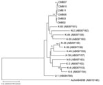
The bovine kobuvirus was detected by the RT-PCR screening method in 6 (8.3%) of the 72 fecal specimens collected. The partial 3D regions of all 6 bovine kobuviruses exhibited highly conserved sequences of 99.3%–100% nucleotide and 100% amino acid identities to each other. In addition, by searching for the closest sequences in the databank, we found that these sequences were closely related to all 13 bovine kobuvirus reference strains available in the GenBank database (U-1, K-35, K-36, K-49, K-38, N-5, K-3, K-4, K-6, K-60, N-2, K-44, and K-55); nucleotide and amino acid sequence identities ranged from 91.0% to 95.0% and 97.4% to 99.4%, respectively. Phylogenetic analysis of partial 3D nucleotide sequences of our bovine kobuvirus strains, together with those of all published bovine kobuvirus reference strains available in the GenBank database, is shown in the Figure. The Aichi virus standard strain was included in the tree as an outlier virus. The phylogenetic tree confirmed that all 6 virus strains belonged to bovine kobuvirus and formed a tight cluster in a monophyletic branch with other published bovine kobuvirus reference strains, but they were distantly related to the Aichi virus standard strain.
Detection and characterization of bovine kobuvirus strains from different geographic areas are important for understanding the worldwide distribution, heterogeneity, and association of bovine kobuvirus with enteric disease in cattle. Our findings indicate the role of kobuviruses in diarrheal disease in cattle and provide additional information on their relationship to bovine kobuviruses reported previously.
Acknowledgment
This research was supported by the Core University System Exchange Program under the Japan Society for the Promotion of Science, coordinated by the Graduate School of Medicine, the University of Tokyo, Japan, and Mahidol University, Thailand. The study was also supported in part by the Research Fund from the Faculty of Medicine, Chiang Mai University, Thailand.
References
- Yamashita T, Ito M, Kabashima Y, Tsuzuki H, Fujiura A, Sakae K. Isolation and characterization of a new species of kobuvirus associated with cattle. J Gen Virol. 2003;84:3069–77. DOIPubMedGoogle Scholar
- Racaniello VR. Picornaviridae: the viruses and their replication. In: Knipe DM and Howley PM, editors. Fields virology, vol. 1. 5th ed. Philadelphia: Lippincott Williams & Wilkins; 2006. p. 795–838.
- Oh DY, Silva PA, Hauroeder B, Diedrich S, Cardoso DD, Schreier E. Molecular characterization of the first Aichi viruses isolated in Europe and in South America. Arch Virol. 2006;151:1199–206. DOIPubMedGoogle Scholar
- Pham NT, Khamrin P, Nguyen TA, Kanti DS, Phan TG, Okitsu S, Isolation and molecular characterization of Aichi viruses from fecal specimens collected in Japan, Bangladesh, Thailand, and Vietnam. J Clin Microbiol. 2007;45:2287–8. DOIPubMedGoogle Scholar
- Yamashita T, Sakae K, Kobayashi S, Ishihara Y, Miyake T, Mubina A, Isolation of cytopathic small round virus (Aichi virus) from Pakistani children and Japanese travelers from Southeast Asia. Microbiol Immunol. 1995;39:433–5.PubMedGoogle Scholar
- Altschul SF, Gish W, Miller W, Myers EW, Lipman DJ. Basic local alignment search tool. J Mol Biol. 1990;215:403–10.PubMedGoogle Scholar
- Kumar S, Tamura K, Nei M. MEGA3: integrated software for molecular evolutionary genetics analysis and sequence alignment. Brief Bioinform. 2004;5:150–63. DOIPubMedGoogle Scholar
Figure
Cite This ArticleRelated Links
Table of Contents – Volume 14, Number 6—June 2008
| EID Search Options |
|---|
|
|
|
|
|
|

Please use the form below to submit correspondence to the authors or contact them at the following address:
Hiroshi Ushijima, Department of Developmental Medical Sciences, Institute of International Health, Graduate School of Medicine, University of Tokyo, 7-3-1 Hongo, Bunkyo-ku, Tokyo 113-0033, Japan;
Top