Volume 15, Number 1—January 2009
Dispatch
Imported Case of Poliomyelitis, Melbourne, Australia, 2007
Andrew J. Stewardson , Jason A. Roberts, Carolyn L. Beckett, Hayden T. Prime, Poh-Sien Loh, Bruce R. Thorley, and John R. Daffy
Figure 1
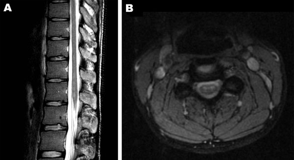
Figure 1. A) Sagittal image of the conus and B) coronal image of the cervical cord, demonstrating increased signal on T2 weighted sequences in the region of the anterior horns. There was no enhancement with contrast.
Page created: December 06, 2010
Page updated: December 06, 2010
Page reviewed: December 06, 2010
The conclusions, findings, and opinions expressed by authors contributing to this journal do not necessarily reflect the official position of the U.S. Department of Health and Human Services, the Public Health Service, the Centers for Disease Control and Prevention, or the authors' affiliated institutions. Use of trade names is for identification only and does not imply endorsement by any of the groups named above.