Volume 15, Number 2—February 2009
Dispatch
Enteroviruses in Patients with Acute Encephalitis, Uttar Pradesh, India
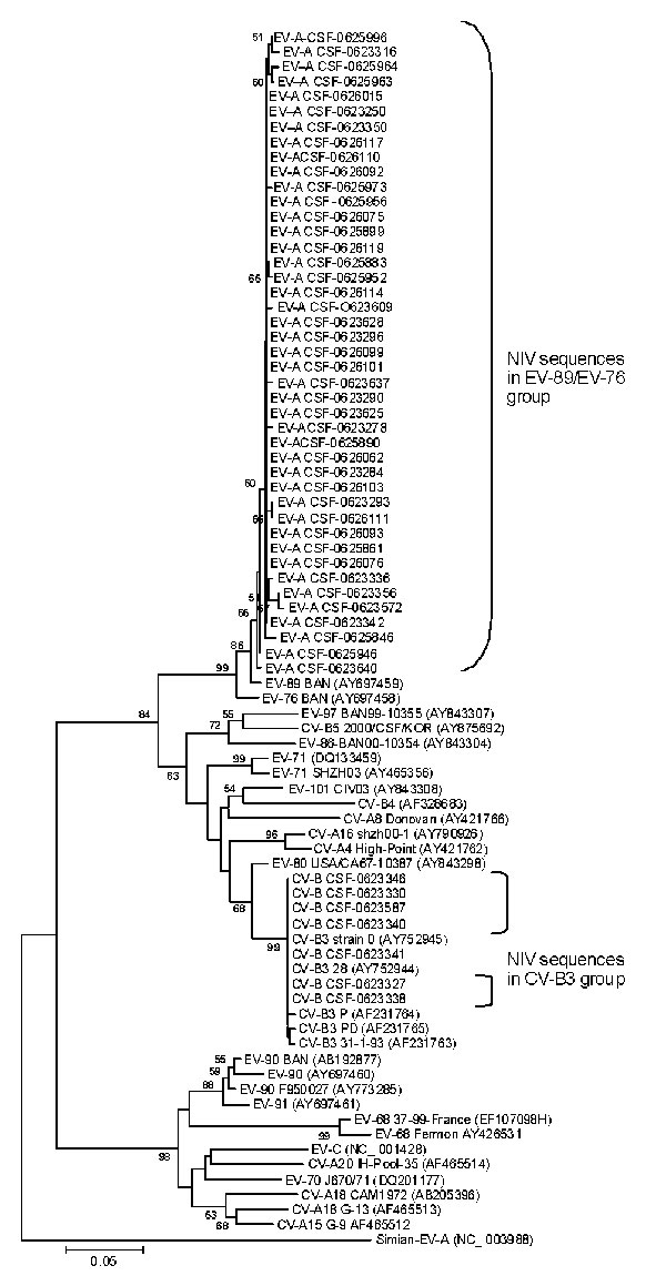
Figure 1

Figure 1. Phylogenetic tree based on partial 5’ noncoding region sequences of enterovirus (EV) genome detected in cerebrospinal fluid samples from encephalitis patients. Specimens are identified by repository serial numbers obtained from the National Institute of Virology (NIV), Pune, India. GenBank accession nos. EU672893–EU762967 indicate the nucleotide sequences of EV strains of the present study. Scale bar indicates nucleotide substitutions per site. EV, enterovirus; CSF, cerebrospinal fluid; CV-A, coxsackie virus A; CV-B, coxsackie virus B; HEV, human enterovirus.
Page created: December 08, 2010
Page updated: December 08, 2010
Page reviewed: December 08, 2010
The conclusions, findings, and opinions expressed by authors contributing to this journal do not necessarily reflect the official position of the U.S. Department of Health and Human Services, the Public Health Service, the Centers for Disease Control and Prevention, or the authors' affiliated institutions. Use of trade names is for identification only and does not imply endorsement by any of the groups named above.