Skip directly to site content Skip directly to page options Skip directly to A-Z link Skip directly to A-Z link Skip directly to A-Z link
Volume 15, Number 2—February 2009
Research

Bacterial Phenotype Variants in Group B Streptococcal Toxic Shock Syndrome1

Parham Sendi2Comments to Author , Linda Johansson, Samira Dahesh, Nina M. Van Sorge, Jessica Darenberg, Mari Norgren, Jan Sjölin, Victor Nizet, and Anna Norrby-Teglund
Author affiliations: Karolinska Institutet, Stockholm, Sweden (P. Sendi, L. Johansson, A. Norrby-Teglund); Basel University Medical Clinic, Liestal, Switzerland (P. Sendi); University of California San Diego, La Jolla, California, USA (S. Dahesh, N.M. Van Sorge, V. Nizet); Swedish Institute for Infectious Disease Control, Stockholm (J. Darenberg); Umeå University, Umeå, Sweden (M. Norgren); Uppsala University, Uppsala, Sweden (J. Sjölin)

Main Article

Figure 7

A) Human whole-blood killing assay after 3 h incubation, using 100 CFU bacteria in 100 μL phosphate-buffered saline (PBS) and 300 μL blood. Error bars indicate SEM. B) 100 CFU bacteria in 100 μL PBS and 1,000 μL blood. Survival index is calculated as follows: (CFU at the end of the assay)/(CFU at t = 0 h). Horizontal lines indicate the median. C) Opsonophagocytic killing assay after 1 h incubation, using a multiplicity of infection of ≈1:1 (CFU 106/mL: neutrophils 106/mL) and 10% volume serum. E

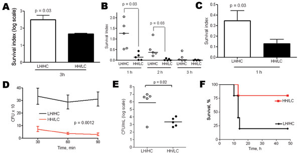
Figure 7. A) Human whole-blood killing assay after 3 h incubation, using 100 CFU bacteria in 100 μL phosphate-buffered saline (PBS) and 300 μL blood. Error bars indicate SEM. B) 100 CFU bacteria in 100 μL PBS and 1,000 μL blood. Survival index is calculated as follows: (CFU at the end of the assay)/(CFU at t = 0 h). Horizontal lines indicate the median. C) Opsonophagocytic killing assay after 1 h incubation, using a multiplicity of infection of ≈1:1 (CFU 106/mL: neutrophils 106/mL) and 10% volume serum. Error bars indicate SEM. D) Intracellular survival assays in neutrophils after 30, 60, and 90 min of extracellular antimicrobial drug exposure. Error bars indicate SEM. E) Murine model for invasive disease (low inoculum). Groups of 5 mice were inoculated intraperitoneally with 6–8 × 10⁶ CFU of low hemolytic (LH)/high encapsulation (HC) or high hemolytic (HH)/low encapsulation (LC) group B streptococcal isolates per mouse. Levels of bacteremia were assessed after 6 h. Horizontal lines indicate the median. F) Kaplan-Meier survival plot.

Main Article

1Part of this work was presented at Lancefield 2008 International Symposium on Streptococci and Streptococcal Diseases, Porto Heli, Greece, June 22–26, 2008.

2Current affiliation: Clinic for Infectious Diseases, University Hospital Bern, Bern, Switzerland.

Page created: December 08, 2010
Page updated: December 08, 2010
Page reviewed: December 08, 2010
The conclusions, findings, and opinions expressed by authors contributing to this journal do not necessarily reflect the official position of the U.S. Department of Health and Human Services, the Public Health Service, the Centers for Disease Control and Prevention, or the authors' affiliated institutions. Use of trade names is for identification only and does not imply endorsement by any of the groups named above.
file_external