Volume 15, Number 2—February 2009
Letter
Mesotherapy-associated Outbreak Caused by Mycobacterium immunogenum
Cite This Article
Citation for Media
To the Editor: Mesotherapy, a procedure for medical and cosmetic treatment, involves use of microinjections of different biologically active substances into the dermis or subcutaneous adipose tissue. This controversial practice is used for spot contouring and anti-aging therapy. Concerns have been raised about mesotherapy complications, such as aseptic subcutaneous necrosis and cutaneous nontuberculous mycobacterial infections. Several rapidly growing mycobacterial species, primarily Mycobacterium fortuitum, M. peregrinum, M. chelonae, M. abscessus, M. simiae, and the newly described M. massiliense, M. bolletii, and M. cosmeticum (1–5), have been reported to cause infections and outbreaks originating from use of contaminated injectable solutions or skin antiseptics during mesotherapy and other invasive cosmetic procedures. We describe a mesotherapy-associated outbreak involving an organism compatible with the novel M. immunogenum that Wilson et al. first described (6).
During September 2006–May 2007, 169 persons underwent mesotherapy at a private aesthetic clinic in the city of Buenos Aires, Argentina. For 28 (17%) skin lesions developed at the injection sites. Patients had been injected during the first 2 months of 2007 with phosphatidylcholine and ampelopsine after receiving a topical antiseptic containing lapyrium chloride from a commercial supplier. The clinic and all the products used in the procedure had been licensed by the national regulatory authority. As soon as the outbreak became evident, the antiseptic solution in use was discarded; no additional cases occurred. By the time the investigation was conducted, these solutions were no longer available for culture.
Nineteen patients were referred to our hospitals. Physical examination found 2–20 nodules 0.5–4 cm in diameter per patient. Lesions were localized on legs (18 patients), buttocks (16), abdomen (1), and forearms (1) and had appeared 7–37 days (median 31 days) after the injection. Occasionally, some nodules produced a serous or purulent discharge, but secretions were not submitted for bacteriologic analysis.
Nodule biopsy was performed for 10 patients, and specimens were sent for histologic and bacteriologic investigations. Histologic examination demonstrated abscesses, areas of fat necrosis, and peripheral fibrous changes. Specimens from 3 patients produced acid-fast bacilli growth in blood agar, mycobacteria growth indicator tube, and Lowenstein-Jensen culture media. When subcultured at 3 different temperatures for identification, all 3 cultures grew preferentially at room temperature rather than at 37°C and 42°C. The isolated mycobacteria were nonpigmented, rapidly growing, and fastidious. The results of biochemical tests produced a hybrid pattern between M. chelonae and M. abscessus. Specifically, the isolates were unable to use citrate as the sole carbon source and to grow in the presence of 5% NaCl.
Figure
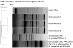
Figure. DNA enterobacterial repetitive intergenic consensus PCR (eric) analysis of rapidly growing mycobacteria isolated from 3 patients in a mesotherapy-associated outbreak, January–February, 2007, Buenos Aires city, Argentina, compared with profiles of epidemiologically...
PCR-restriction analysis (PRA) of a 439-bp segment of the hsp65 gene digested with BstEII and HaeIII was performed at the national reference laboratory for tuberculosis of the Instituto Malbran. Species was assigned according to the PRASITE website (http://app.chuv.ch/prasite). The profile of the 3 isolates fit the pattern of M. immunogenum type 2 as first described by Sampaio et al. (7); the isolates had 325- and 130-bp bands after BstEII digestion and 200-, 70-, 58-, and 55-bp bands after HaeIII digestion. This is our first detection of this particular PRA profile in Argentina since we started systematic hsp65 PRA typing to identify mycobacteria in clinical isolates in 2005. Enterobacterial repetitive intergenic consensus PCR patterns of the 3 isolates were indistinguishable from each other and differed from epidemiologically unrelated clinical isolates of the M. abscessus–M. chelonae group, confirming the clonality of the 3 strains (Figure) (7).
Susceptibility to antimicrobial agents was determined by using standard Clinical and Laboratory Standards Institute broth microdilution method (8). Clarithromycin, ciprofloxacin, cefoxitin, doxycycline, amikacin, tobramycin, and imipenem MIC values for the 3 isolates were <0.125, 1, 32, >32, 32, 16, and 64 µg/mL, respectively. The disk elution method produced similar patterns of activity for the first 4 antimicrobial agents and inconsistent results for the remaining 3.. Patients received a combination of clarithromycin and either ciprofloxacin or levofloxacin for 6–8 months. All 19 cases resolved favorably, although multiple pigmented retractile scars persisted after treatment.
M. immunogenum was identified as the etiologic agent of a variety of hospital-acquired infections, including an outbreak of keratitis, and as the potential cause of hypersensitivity pneumonitis in industrial metal-grinding machinists (6,7,9). This microorganism appears to differ from other members of the M. chelonae–abscessus group by subtle mutations in rpoB, hsp65, the hypervariable region of 16S rRNA, and other housekeeping genes (5–7,9,10). The value of minor polymorphisms might be arguable for defining new species and for clinically managing patients. However, because of its rarity among clinical isolates in our country, the PRA type ascribed to M. immunogenum proved to be a useful epidemiologic marker to investigate this outbreak.
References
- Centers for Disease Control and Prevention. Outbreak of mesotherapy-associated skin reactions—District of Columbia area, January–February 2005. MMWR Morb Mortal Wkly Rep. 2005;54:1127–30.PubMedGoogle Scholar
- Cooksey RC, de Waard JH, Yakrus MA, Rivera I, Chopite M, Toney SR, Mycobacterium cosmeticum sp. nov., a novel rapidly growing species isolated from a cosmetic infection and from a nail salon. Int J Syst Evol Microbiol. 2004;54:2385–91. DOIPubMedGoogle Scholar
- Rivera-Olivero IA, Guevara A, Escalona A, Oliver M, Pérez-Alfonzo R, Piquero J, Soft-tissue infections due to non-tuberculous mycobacteria following mesotherapy. What is the price of beauty [in Spanish]. Enferm Infecc Microbiol Clin. 2006;24:302–6. DOIPubMedGoogle Scholar
- Munayco CV, Grijalva CG, Culqui DR, Bolarte JL, Suárez Ognio LA, Quispe N, Outbreak of persistent cutaneous abscesses due to Mycobacterium chelonae after mesotherapy sessions, Lima, Peru. Rev Saude Publica. 2008;42:146–9. DOIPubMedGoogle Scholar
- Viana-Niero C, Lima KV, Lopes ML, Rabello MC, Marsola LR, Brilhante VC, Molecular characterization of Mycobacterium massiliense and Mycobacterium bolletii in isolates collected from outbreaks of infections after laparoscopic surgeries and cosmetic procedures. J Clin Microbiol. 2008;46:850–5. DOIPubMedGoogle Scholar
- Wilson RW, Steingrube VA, Böttger EC, Springer B, Brown-Elliott BA, Vincent V, Mycobacterium immunogenum sp. nov., a novel species related to Mycobacterium abscessus and associated with clinical disease, pseudo-outbreaks, and contaminated metalworking fluids: an international cooperative study on mycobacterial taxonomy. Int J Syst Evol Microbiol. 2001;51:1751–64.PubMedGoogle Scholar
- Sampaio JL, Junior DN, de Freitas D, Höfling-Lima AL, Miyashiro K, Alberto FL, An outbreak of keratitis caused by Mycobacterium immunogenum. J Clin Microbiol. 2006;44:3201–7. DOIPubMedGoogle Scholar
- Clinical and Laboratory Standards Institute. Susceptibility testing of mycobacteria, nocardiae, and other aerobic actinomycetes: approved standard [CLSI document M24-A]. Wayne (PA): The Institute; 2003.
- Wallace RJ Jr, Zhang Y, Wilson RW, Mann L, Rossmoore H. Presence of a single genotype of the newly described species Mycobacterium immunogenum in industrial metalworking fluids associated with hypersensitivity pneumonitis. Appl Environ Microbiol. 2002;68:5580–4.PubMedGoogle Scholar
- Adékambi T, Drancourt M. Dissection of phylogenetic relationships among 19 rapidly growing Mycobacterium species by 16S rRNA, hsp65, sodA, recA and rpoB gene sequencing. Int J Syst Evol Microbiol. 2004;54:2095–105. DOIPubMedGoogle Scholar
Figure
Cite This ArticleRelated Links
Table of Contents – Volume 15, Number 2—February 2009
| EID Search Options |
|---|
|
|
|
|
|
|
Please use the form below to submit correspondence to the authors or contact them at the following address:
Domingo J. Palmero, N. Videla 559, (1424) Buenos Aires, Argentina;
Top