Volume 15, Number 4—April 2009
THEME ISSUE
The Amazon Region
Research
Human Febrile Illness Caused by Encephalomyocarditis Virus Infection, Peru
Figure 1
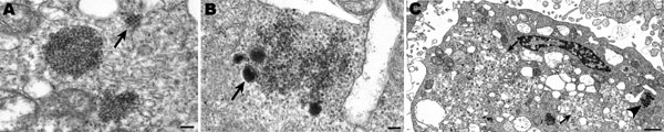
Figure 1. Ultrastructural morphologic features of cardiovirus-infected Vero E6 cells. A) Collections of picornavirus particles, some arranged in a paracrystalline array (arrow). Scale bar = 100 nm. B) Higher magnification of area pointed to by arrowhead in panel C showing condensed material (arrow) at periphery of a viral cluster. Scale bar = 100 nm. C) Cardiovirus-infected cell, showing membrane proliferation and vesiculation (arrows). Scale bar = 1 μm.
Page created: December 10, 2010
Page updated: December 10, 2010
Page reviewed: December 10, 2010
The conclusions, findings, and opinions expressed by authors contributing to this journal do not necessarily reflect the official position of the U.S. Department of Health and Human Services, the Public Health Service, the Centers for Disease Control and Prevention, or the authors' affiliated institutions. Use of trade names is for identification only and does not imply endorsement by any of the groups named above.