Volume 16, Number 11—November 2010
Research
Lymphotropism of Merkel Cell Polyomavirus Infection, Nova Scotia, Canada
Figure

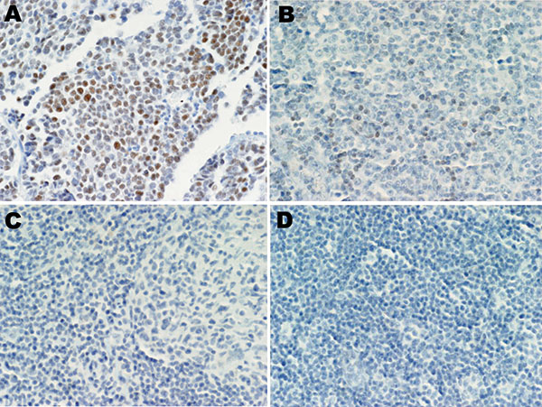
Figure. Merkel cell polyomavirus (MCPyV) large T-antigen (T-ag) expression in human tissues. A) Merkel cell carcinoma stained with CM2B4 antibody as a positive control; MCPyV T-ag was detected. B) Expression of MCPyV T-ag in small lymphocytes in an MCPyV DNA–positive angioimmunoblastic T-cell lymphoma, stained with CM2B4. C) MCPyV DNA–positive reactive lymphoid hyperplasia sample reacted with CM2B4; no T-ag was detected. D) MCPyV DNA-negative chronic lymphocytic leukemia/small lymphocytic lymphoma sample stained with CM2B4; no T-ag was detected. Original magnification ×40.
Page created: March 08, 2011
Page updated: March 08, 2011
Page reviewed: March 08, 2011
The conclusions, findings, and opinions expressed by authors contributing to this journal do not necessarily reflect the official position of the U.S. Department of Health and Human Services, the Public Health Service, the Centers for Disease Control and Prevention, or the authors' affiliated institutions. Use of trade names is for identification only and does not imply endorsement by any of the groups named above.