Volume 16, Number 4—April 2010
Research
Influenza A Strain-Dependent Pathogenesis in Fatal H1N1 and H5N1 Subtype Infections of Mice
Figure 6

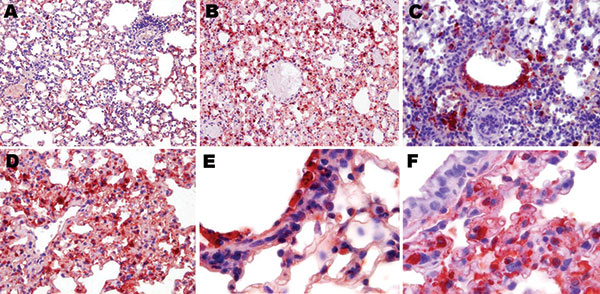
Figure 6. Topologic distribution of influenza antigens in the lungs of mice infected with influenza virus A subtype H1N1 and H5N1 strains at endpoint (antinucleoprotein immunohistochemical staining). A) Subtype H1N1 and B) subtype H5N1, both showing diffusely distributed positive staining of numerous pneumocytes and alveolar macrophages (original magnification ×100). C) Subtype H1N1, showing antigens massively present in the remaining non-desquamated airway epithelial cells (original magnification ×400); viral amplification in type I and type II pneumocytes is far more intense and widespread 4 days after inoculation of the subtype H5N1 virus (D) than 7 days after inoculation of the subtype H1N1 virus (original magnification ×400). E) Desquamated, necrotic, and intensely virus-positive airway epithelial cells in a terminal bronchiole and adjacent alveoli of a mouse infected with subtype H1N1, compared with F) uninfected, intact airway epithelial cells in a terminal bronchiole and adjacent alveoli of a mouse infected with subtype H5N1, illustrating the different pneumotropism of the 2 viruses (original magnification ×1,000). Conversely, the density of virus-positive cells in the lung/alveoli is higher after inoculation of the subtype H5N1 strain (Mayer hematoxylin counterstain).