Volume 16, Number 4—April 2010
Research
Influenza A Strain-Dependent Pathogenesis in Fatal H1N1 and H5N1 Subtype Infections of Mice
Mutien-Marie Garigliany, Adélite Habyarimana, Bénédicte Lambrecht, Els Van de Paar, Anne Cornet, Thierry van den Berg, and Daniel Desmecht
Figure 7
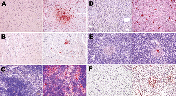
Figure 7. Topologic distribution of antigens in mice infected with influenza A virus subtype H1N1 at day 7 postinfection (left columns) and subtype H5N1 at day 4 postinfection (right columns) in various nonrespiratory organs. A) Glial cells (mostly oligodendrocytes); B) cardiomyocytes; C) spleen macrophages; D) hepatocytes; E) islets of Langerhans cells in the pancreas; and F) adipocytes. Bright virus-positive staining can be seen in subtype H5N1–infected mice (antinucleoprotein immunohistochemical staining), while absence of any staining can be seen in subtype H1N1–infected mice (Mayer hematoxylin counterstain). Original magnification ×100.
Page created: December 28, 2010
Page updated: December 28, 2010
Page reviewed: December 28, 2010
The conclusions, findings, and opinions expressed by authors contributing to this journal do not necessarily reflect the official position of the U.S. Department of Health and Human Services, the Public Health Service, the Centers for Disease Control and Prevention, or the authors' affiliated institutions. Use of trade names is for identification only and does not imply endorsement by any of the groups named above.