Volume 16, Number 9—September 2010
Letter
Invasive Klebsiella pneumoniae Infections, California, USA
Robert McCabe , Larry Lambert, and Brad Frazee
Figure
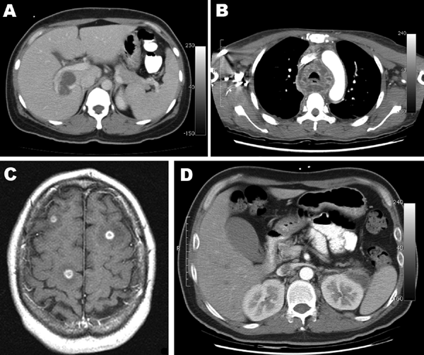
Figure. A) Computed tomography (CT) scan of the abdomen showing a liver abscess adjacent to the portal vein. B) CT scan of the chest at the level of the aortic arch showing mediastinum abscesses surrounding the trachea. C) Brain magnetic resonance imaging (T1 weighted, spin echo, with contrast) showing multiple intracerebral abscesses (smooth ring-enhancing lesions with surrounding vasogenic edema). D) CT scan of the abdomen of patient from panel C, showing a left perinephric abscess and thrombus.
Page created: August 28, 2011
Page updated: August 28, 2011
Page reviewed: August 28, 2011
The conclusions, findings, and opinions expressed by authors contributing to this journal do not necessarily reflect the official position of the U.S. Department of Health and Human Services, the Public Health Service, the Centers for Disease Control and Prevention, or the authors' affiliated institutions. Use of trade names is for identification only and does not imply endorsement by any of the groups named above.