Volume 17, Number 12—December 2011
Research
Isolation of Prion with BSE Properties from Farmed Goat
Figure 1
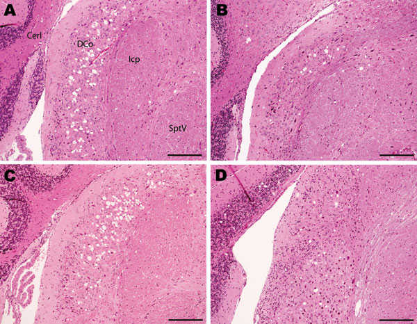
Figure 1. Histopathologic analysis of cochlear nuclei from host-encoded prion protein (PrP)-a mice (C57/BL6) inoculated with (A) fixed material from the suspected case, (B) fixed material from experimental goat bovine spongiform encephalopathy (BSE), (C) unfixed material from experimental sheep BSE, and (D) fixed material from experimental goat scrapie. The BSE-challenged mice (A–C) show confluent vacuolation in the dorsal cochlear nucleus that extends ventrally with increasing lesion severity. Even in mild cases (B) this lesion can be distinguished from the low-frequency randomly dispersed vacuoles observed in scrapie (D). Note the unaffected nature of the lesion between fixed (A and B) and unfixed (C) samples. Cerl, cerebellum; DCo, dorsal cochlear nucleus; Icp, inferior cerebellar peduncle; SptV, spinal tract of the trigeminal nerve. Scale bars = 200 μm.