Volume 17, Number 12—December 2011
Research
Isolation of Prion with BSE Properties from Farmed Goat
Figure 5
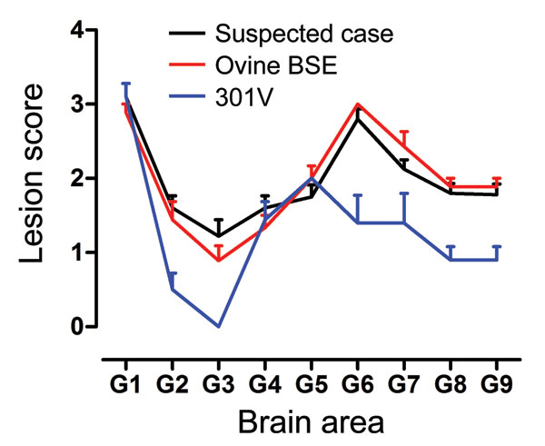
Figure 5. Lesion profiles from VM mice after second passage of the suspected case, serial passage of an ovine bovine spongiform encephalopathy (BSE) source, and a 301V control. Profiles were made on the basis of the lesion score, which is the quantification of transmissible spongiform encephalopathy–specific vacuolation in 9 neuroanatomical gray matter areas: G1, dorsal medulla nuclei; G2, cerebellar cortex of the folia including the granular layer, adjacent to the fourth ventricle; G3, cortex of the superior colliculus; G4, hypothalamus; G5, thalamus; G6, hippocampus; G7, septal nuclei of the paraterminal body; G8, cerebral cortex (at the level of G4 and G5); G9, cerebral cortex (at the level of G7). At least 9 clinically and histopathologically positive mice contributed to each profile. Error bars indicate SEM.