Volume 17, Number 12—December 2011
Research
Isolation of Prion with BSE Properties from Farmed Goat
Figure 2
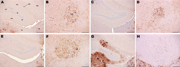
Figure 2. Immunohistochemical analysis of brains of host-encoded prion protein (PrP)-a mice (RIII) inoculated with (A and B) fixed material from the goat with suspected bovine spongiform encephalopathy (BSE), (C and D) fixed material from experimental goat BSE, (E and F) unfixed material from experimental sheep BSE, and (G and H) fixed material from experimental goat scrapie. No PrPSc was detected in the molecular layer of the dentate gyrus in the suspected case (A) and the BSE controls (C and E); in the scrapie control (G) the same area is heavily affected. In the red nucleus, small PrPSc aggregates were observed in the suspected case (B) and in the BSE controls (D and F), whereas the same nucleus seem to be unaffected in the scrapie control despite evident PrPSc deposits in the surrounding area. Hip, hippocampus; Hif, hippocampal fissure; Hb, habenular nuclei; RdN, red nucleus; Th, thalamus; gr, mo, and po, granular, molecular, and polymorph layers, respectively, of the dentate gyrus. Scale bars = 200 μm.