Volume 17, Number 7—July 2011
Research
Asian Lineage of Peste des Petits Ruminants Virus, Africa
Cite This Article
Citation for Media
Abstract
Interest in peste des petits ruminants virus (PPRV) has been stimulated by recent changes in its host and geographic distribution. For this study, biological specimens were collected from camels, sheep, and goats clinically suspected of having PPRV infection in Sudan during 2000–2009 and from sheep soon after the first reported outbreaks in Morocco in 2008. Reverse transcription PCR analysis confirmed the wide distribution of PPRV throughout Sudan and spread of the virus in Morocco. Molecular typing of 32 samples positive for PPRV provided strong evidence of the introduction and broad spread of Asian lineage IV. This lineage was defined further by 2 subclusters; one consisted of camel and goat isolates and some of the sheep isolates, while the other contained only sheep isolates, a finding with suggests a genetic bias according to the host. This study provides evidence of the recent spread of PPRV lineage IV in Africa.
Peste des petits ruminants virus (PPRV) belongs to the genus Morbillivirus, in the family Paramyxoviridae. Like other members of the same genus, such as rinderpest virus, Canine distemper virus, Measles virus, and marine mammal viruses, PPRV is highly pathogenic for its natural hosts (1). As consistently reported by the World Organisation for Animal Health, PPRV causes high death rates in livestock. It has a major economic effect, particularly in the intertropical regions of Africa, on the Arabian Peninsula and in the Middle East and Asia (2–5). The main signs of the acute form of the disease are high fever, mouth ulceration, diarrhea, and pneumonia.
Although rinderpest virus was eradicated after intensive vaccination campaigns in the last quarter of the 20th century (6), PPRV has continued to spread in Africa and Asia. In East Africa, outbreaks occurred in 2007 in Uganda and Kenya and in 2009 in Tanzania (7); in North Africa, an outbreak occurred in 2008 in Morocco. Genotypic classification of PPRV has identified 4 lineages and appears to be an efficient tool to survey virus spread worldwide. Genetic variability is based on partial sequencing of the fusion (F) protein gene (8,9) and of the 3′ end of the nucleoprotein (N) gene (10). Both are well conserved genes with ≈10% nt mean variability between the most distantly related sequences (11); this variability can exceed 30% on some parts of the sequence. However, because the N gene is the most abundantly transcribed virus gene, sensitivity is better achieved with N mRNAs (12). Viruses of lineage I and II are restricted to western and central Africa; lineage III is common to eastern Africa and the southern part of the Middle East. In Asia, only viruses of lineage IV have been detected.
In Sudan, continuous outbreaks of PPRV have occurred for >30 years, mainly in sheep and goats (2,13). Although PPRV infection is well documented in small ruminants, data are rare for other species, such as cattle, buffalo, and camels. In 1997, a PPRV was isolated from pathologic samples collected during a rinderpest-like disease outbreak among buffalo in India (14). In September 2004, outbreaks of PPRV in Sudan affected both sheep and camels (15,16). In camels, a respiratory syndrome was the prominent disease characteristic observed, resembling a previous case reported in Ethiopia during 1995–1996 (17).
More recently, in summer 2008, Morocco reported outbreaks of PPRV for the first time (18). Because PPRV needs close contact for transmission, this new epizootic was likely the result of introduction of the virus into North Africa through the movement of live infected animals. To characterize PPRV strains identified in Sudan during 2000–2009 and in Morocco during the extensive 2008 outbreaks, a phylogenetic analysis was carried out on wild-type PPRV sequences obtained from biological samples collected from sheep, goats, and camels in these 2 countries.
Biological Materials
Biological specimens were collected in different parts of Sudan during 2000–2009 from camels, sheep, and goats that showed clinical signs of PPRV. Additional camel specimens were collected after outbreaks associated with high death rates were reported from the Kassala Region in 2004. A total of 80 field samples, including lung, liver, and spleen, were obtained from 49 camels, 26 sheep, and 5 goats with PPRV-like clinical signs. For most of the animals, virus detection and identification were performed directly on the tissue samples collected; however, virus isolation in tissue culture was attempted for some samples. A historical PPRV strain isolated in Sudan in 1971, the Gedarif PPRV strain, was included in this study.
In Morocco, a total of 36 samples were collected from sheep displaying PPRV signs during the 2008 outbreaks. These included oral and ocular swabs and mesenteric lymph nodes, spleen, lung, and whole blood samples.
Laboratory Investigations
Sample aliquots first were screened for PPRV antigen detection by immunocapture ELISA (ICE test; Biological Diagnostics Supplies, Ltd., Dreghorn, Scotland). A second aliquot was used for viral RNA extraction and virus isolation in tissue culture. Reverse transcription PCR (RT-PCR) amplified the N protein gene directly from tissue samples by using a set of pan-morbillivirus primers as described by Kwiatek et al. (10).
For the sequencing of the N gene, the 3′ 351 nt were obtained by employing primers NP3 and NP4 (19) and through a modification of the initial protocol by using a 1-step method (OneStep RT-PCR mix; QIAGEN, Courtaboeuf, France). For the F gene, 11 samples were selected from samples that previously tested positive by N amplification. For the specific and sensitive detection of PPRV from these clinical field samples, the initial method developed by Forsyth et al. in 1995 (8) was modified by using an additional pair of primer F1/FPPRrev in combination with F1/F2 in a nested PCR. Sequences positions of these primers were 5′-ATCACAGTGTTAAAGCCTGTAGAGG-3′ (PPRV-F1: 777 ± 801), 5′-GAGACTGAGTTTGTGACCTACAAGC-3′ (PPRV-F2: 1124 ± 1148) and 5′-ATATTAATGTGACAAGCCCTAGGGA-3′ (FPPRrev: 2055 ± 2079). The final amplicon was obtained at the expected length of 372 nt after 35 cycles. We analyzed 10 μL of the amplified products by electrophoresis on 1.5% agarose gel. For all positive results, 40 μL of the final product was used directly for sequencing (GATC Biotech, Constance, Germany). All sequences were deposited in GenBank under accession nos. HQ131917– HQ131958. FPPRrev and the study of the target sites of primers F1 and F2 (8) were designed by a comparative sequence analysis of various morbillivirus F sequences from the full genomes available in GenBank (accession nos. X74443, EU267273, EU267274, AY560591, FJ905304, and AJ849636).
Sequence Data, Alignment, and Phylogenetic Analysis
Sequencing reactions were performed by GATC Biotech. Each nucleic acid segment on the N and F genes of the Sudan and Morocco strains was aligned with other sequences from PPRV maintained in the database or retrieved from GenBank (Table 1) (10,20–22). Multiple alignments of 255 and 322 nt of the N and F genes, respectively, were made by using the ClustalW program (www.ebi.ac.uk/clustalw). Phylogenetic analysis was carried out by using the criterion of neighborhood based on the principle of parsimony (23). Dissimilarities and distances between the sequences first were determined with Darwin software (24). Tree construction was based on the unweighted neighbor-joining method proposed by Gascuel (25). Trees were generated with the TreeConMATRIXW program of Darwin (26). Bootstrap confidence intervals were calculated on 1,000 replicates.
Virus Isolation
For the samples from Sudan, isolation was successful on MDBK cells with lung samples from Cam_8, Cam_169, Cam_318, Ov_Soba, and Ov_Al Azaza, collected from 3 camels and 2 sheep, respectively. For the samples from Morocco, PPRV Morocco_08_02 virus was isolated successfully from a sheep lung sample after infection of Vero.DogSLAMtag cells (27). These isolates were sequenced and compared as described above.
Detection of PPRV by N gene RT-PCR
A total of 80 animals from Sudan that initially tested positive in the ICE test were analyzed further by using RT-PCR. Results for 64 animals were positive, including 21 sheep (80.8%), 5 goats (100%), and 38 camels (77.6%) drawn from all of the regions studied: Khartoum, Blue Nile, Northern Sudan, Kassala, Kordofan, and Darfur (Table 2). Of the 36 samples tested from Morocco, 16 yielded a positive RT-PCR.
Characterization of Strains Involved in the Infection of Sheep, Goats, and Camels in Sudan, and in Sheep in Morocco
Of the tissue samples tested, lung and lymph nodes were the most suitable for sequencing because they had the highest viral load, thereby yielding a sufficient amount of PCR product. Sequence also was obtained from several virus isolates, including the historical Gedarif isolate from 1971. The partial N gene sequences were obtained for 26 of the 64 samples from Sudan and 6 of the 16 samples from Morocco. Sequences were aligned with an extended set of PPRV isolate sequences that either were in the database or were retrieved from GenBank where they were described by Kwiatek et al. (10) and Banyard et al. (20).
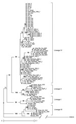
In contrast to what would have been expected for isolates from eastern Africa, most of the PPRV strains collected in Sudan during 2000–2009 were clustered in lineage IV (24); only a few remained in lineage III (2). Molecular typing also showed for the first time the presence of PPRV lineage IV in Morocco (Figure 1).
Lineage IV isolates from Sudan could be further distinguished into 2 clusters. The grouping of the sequences within the clusters was supported by bootstrap percentages >70%. In the first group, 17 sequences from all camel and goat isolates and some sheep isolates (camel Sudan Cam_1, Cam_3, Cam_8, Cam_169, Cam_223, Cam_264, Cam_268, Cam_304, Cam_330, Cam_352, Cam_318; sheep Ov_1, Ov_140, Ov_25, Ov_39300; goat Cap_9, Cap_1) showed 100% identity and matched with Saudi Arabia_1999_7 strain. Sheep Sudan Ov_41 differed from the previous sequences by 2 nt at position 68 (C to T) and 142 (A to G). The divergence of this cluster with consensus IV ranged from 1.2% (Cam_318) to 2% (Ov_41). Furthermore, the Saudi Arabia cluster was related closely to the 6 strains from Morocco collected from the 2008 outbreak, the closest differing by 4 nt.
A separate cluster containing only sheep samples collected during 2000–2008 (sheep Sudan Ov_10033, Ov_10034, Ov_23, Ov_Soba, Ov_Al Azaza) matched with 2 strains from central Africa (Cameroon_1997 [CAMER_1997] and Central African Republic_2004 [CAR_2004]) (20). This cluster was less homogeneous than the cluster previously described; nucleotide variations from the lineage IV consensus sequence ranged from 1.6% (Ov_Al Azaza) to 3.9% (Ov_23).
Only 2 isolates collected at the start of the study (in mid-2000) from sheep in western and eastern Sudan fell into lineage III. Sequences of these 2 field isolates remained similar to the historical strains of Sinnar 72 that was isolated 40 years earlier from the Blue Nile region (2), and with the Gedarif PPRV strain that was isolated from sheep in 1971 (13). In addition, these isolate sequences were close to an Omani strain, Ibri_1983 (28) and a United Arab Emirates strain, Dorcas_1986 (29), but they differed from these strains with a nucleotide variation of 4.7%.
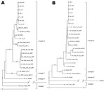
Because the dataset within the Saudi Arabia cluster was highly homogeneous, we compared an N phylogenetic tree (Figure 2, panel A) with an F phylogenetic tree (Figure 2, panel B) on a panel of viruses. These viruses were selected from the 2 lineage IV clusters defined in Figure 1 and from lineage III with additional PPRV from Asia, for which both gene sequences were available (21,22). The F phylogenetic tree confirmed the high homology within the Saudi Arabia cluster, and the cluster’s closeness to strains from Morocco. It also allowed for assignment of strains from Sudan to the 2 described clusters represented by the Arabia_1999_7 and the Central Africa strains.
Unlike the N gene amplifications, amplications with F-specific primers (8) resulted in a number of negative samples by RT-PCR because of mismatches occurring mainly in the F2 reverse primer (Table 3). To circumvent this problem, another reverse primer (FPPRrev) was designed that matched with all lineages.
The sequences and the corresponding lineage classification generated in this study were collated to field information. Occurrence among species of the different lineages show that most of the isolates belong to lineage IV; all camel isolates grouped in the cluster Saudi Arabia, whereas only sheep isolates were found within the other cluster, Central Africa. Only sheep isolates were found in lineage III (Table 4). Geographic and times distribution according to animal species and lineage in Sudan during 2000–2009 show that lineage IV was circulating as early as mid-2000 and that progressive substitution of lineage III in a large zone encompassing the eastern, northern, Blue Nile, and Khartoum regions has taken place since this date (Figure 3).
This study presents findings from PPRV surveillance in Sudan over a 10-year period and the results of tests conducted in Sudan and in Morocco during Morocco’s 2008 outbreaks (18). Analysis of 80 samples confirmed the wide distribution of PPRV throughout Sudan, which has been known since 1971 (2,13,15). Genetic characterization of 26 samples positive for PPRV also provided strong evidence of the introduction and spread of Asia PPRV lineage IV in the country. Surprisingly, the number of samples containing the indigenous lineage III decreased dramatically during the study.
Viruses collected in Morocco also were classified as lineage IV. The origin of the outbreaks in Morocco remains unknown, although Ayari-Fakhfakh et al. (30) recently reported a PPRV seroprevalence in Tunisia of up to 7.45%, indicating that the virus is present across a wider area of northern Africa.
Analysis of the N and F genes showed that 2 clusters were identified in strains from Sudan: 1 related to a strain from Saudi Arabia, the other related to central Africa viruses. The 6 strains from Morocco collected during the 2008 outbreaks were related closely to the Saudi Arabia cluster.
Within the Saudi Arabia cluster, a remarkable genetic stability over a decade was observed; notably, all camel isolates fell into this cluster. The variability and nucleotide sequence was slightly higher for isolates in the central Africa cluster, within which only sheep were found. The inconsistent low genetic variability of isolates from Sudan may be a consequence of a species bias because of the limited contact between camels and sheep. When replicated in a single host that has a limited exposure to new variants, the viral genome thus may remain highly conservative. This hypothesis was verified previously for the measles virus over a shorter period (1997–2000). For the measles virus, it appears that the lower the level of circulation, the higher the sequence conservation (31).
Camels were not regarded as possible hosts for PPRV until 1992, when a number of authors reported PPRV seroconversion in these animals (32–36). The first documented outbreak of PPRV in camels, reported from Ethiopia in 1996, consisted of highly contagious respiratory syndromes with high illness rates but low death rates (17,37). The causative agent was confirmed to be a lineage III PPRV (A. Diallo, unpub. data). This present study confirmed the etiology of the disease in camels through the virologic and epidemiologic investigations and the isolation of PPRV. Surveillance of camels furthermore allowed the virus to be detected in consecutive outbreaks in Kassala, eastern Sudan (2004); Atbara, northern Sudan (2005); and Tambool, Blue Nile region, Sudan (2007) (16). Viruses from the same cluster also were recovered from sick sheep and goats in a large zone encompassing the eastern, northern, Blue Nile, and Khartoum regions as early as mid-2000. Under extensive pastoral farming conditions in these regions, camels may have served as a bridge with areas of northern Africa and contributed to the spread of a camel-derived strain of lineage IV, as seen in Morocco. In contrast, none of the camel and goat isolates were distributed in the central Africa cluster, although viruses of this cluster were cocirculating with camel viruses during the same period and in the same areas of northern Sudan, Khartoum, and Blue Nile.
The reason that isolates of lineage IV became predominant in Sudan, progressively replacing lineage III viruses, during the last decade remains unclear. Lineage IV has been present in Asia and in part of the Middle East for a long time, probably as low virulent strains (5). However, a constant rise of disease incidence recently has been associated with this lineage and suggests increased virulence (3). A virulent lineage IV strain may have been introduced in Africa during the 1990s, resulting in outbreaks in both camels and small ruminants. In parallel, rinderpest virus control and eradication may have favored the decline of cross-immunity in small ruminants and their increased risk for PPRV as predicted earlier by Taylor (38).
Mr Kwiatek works in the Morbillivirus group of the emerging and infectious animal diseases unit at CIRAD, where he is in charge of diagnostic activity of the World Animal Health Organisation and FAO Rinderpest and Peste des Petits Ruminants Reference Laboratory. His research interests involve phylogenetic studies and development of diagnostic tools for rinderpest virus and PPRV.
Acknowledgments
We thank staff at the French Embassy of Sudan for their assistance in funding the scholarship of Y.H.A. to Centre International de Recherche Agronomique pour le Développement (CIRAD).
This study was partially supported by grants from the European Union Network of Excellence, EPIZONE (contract no. FOOD-CT-2006-016236).
References
- Gibbs EPJ, Taylor WP, Lawman MPJ, Bryant J. Classification of the peste des petits ruminants virus as the fourth member of the genus Morbillivirus. Intervirology. 1979;11:268–74. DOIPubMedGoogle Scholar
- El Hag Ali B, Taylor WP. Isolation of peste des petits ruminants virus from Sudan. Res Vet Sci. 1984;36:1–4.PubMedGoogle Scholar
- Shaila MS, Shamaki D, Forsyth MA, Diallo A, Kitching RP, Barrett T. Geographic distribution and epidemiology of peste des petits ruminants virus. Virus Res. 1996;43:149–53. DOIPubMedGoogle Scholar
- Taylor WP, Barrett T. Peste de petits ruminants and rinderpest. In: Aitken ID, editor. Diseases of sheep, 4th ed. Oxford: Blackwells Science; 2007. p. 460–8.
- World Organisation for Animal Health. Immediate notifications and follow-up reports. World Animal Health Information Database (WAHID) Interface; 2009 [cited 2009 Jan 27]. http://www.oie.int/wahis/public.php?page=reports_pdf_download
- Forsyth MA, Barrett T. Evaluation of polymerase chain reaction for the detection and characterisation of rinderpest and peste des petits ruminants viruses for epidemiological studies. Virus Res. 1995;39:151–63. DOIPubMedGoogle Scholar
- Ozkul A, Akca Y, Alkan F, Barrett T, Karaoglu T, Dagalp SB, Prevalence, distribution, and host range of peste des petits ruminants virus, Turkey. Emerg Infect Dis. 2002;8:708–12.PubMedGoogle Scholar
- Kwiatek O, Minet C, Grillet C, Hurard C, Carlsson E, Karimov B, Peste des petits ruminants (PPR) outbreak in Tajikistan. J Comp Pathol. 2007;136:111–9. DOIPubMedGoogle Scholar
- Chard LS, Bailey DS, Dash P, Banyard AC, Barrett T. Full genome sequences of two virulent strains of peste-des-petits ruminants virus, the Côte d'Ivoire 1989 and Nigeria 1976 strains. Virus Res. 2008;136:192–7. DOIPubMedGoogle Scholar
- Barrett T, Banyard AC, Diallo A. Molecular biology of the morbilliviruses. In: Barrett T, Pastouret PP, Taylor WP, editors. Rinderpest and peste des petits ruminants virus. London: Academic Press; 2006. p. 31–56.
- Ali B el-H. A natural outbreak of rinderpest involving sheep, goats and cattle in Sudan. [PMID: 4807688]. Bull Epizoot Dis Afr. 1973;12:421–8.
- Govindarajan R, Koteeswaran A, Venugopalan AT, Shyam G, Shaouna S, Shaila MS, Isolation of peste des petits ruminants virus from an outbreak in Indian buffalo (Bubalus bubalis). Vet Rec. 1997;141:573–4. DOIPubMedGoogle Scholar
- Saeed IK, Ali YH, Khalafalla AI, Rahman-Mahasin EA. Current situation of peste des petits ruminants (PPR) in the Sudan. Trop Anim Health Prod. 2010;42:89–93. DOIPubMedGoogle Scholar
- Khalafalla AI, Saeed IK, Ali YH, Abdurrahman MB, Kwiatek O, Libeau G, An outbreak of peste des petits ruminants (PPR) in camels in the Sudan. Acta Trop. 2010;116:161–5. DOIPubMedGoogle Scholar
- Roger F, Guebre Yesus M, Libeau G, Diallo A, Yigezu LM, Yilma T. Detection of antibodies of rinderpest and peste des petits ruminants viruses (Paramyxoviridae, Morbillivirus) during a new epizootic disease in Ethiopian camels (Camelus dromedarius). Rev Med Vet (Toulouse). 2001;152:265–8.
- Sanz-Alvarez J, Diallo A, De La Rocque S, Pinto J, Thevenet S, Lubroth J. Peste des petits ruminants (PPR) in Morocco. EMPRES Watch, 2008 August 1–7 [cited 2009 Jan 27]. ftp://ftp.fao.org/docrep/fao/011/aj120f/aj120f00.pdf
- Couacy-Hymann E, Roger F, Hurard C, Guillou JP, Libeau G, Diallo A. Rapid and sensitive detection of peste des petits ruminants virus by a polymerase chain reaction assay. J Virol Methods. 2002;100:17–25. DOIPubMedGoogle Scholar
- Banyard AC, Parida S, Batten C, Oura C, Kwiatek O, Libeau G. Global distribution of peste des petits ruminants virus and prospects for improved diagnosis and control. J Gen Virol. 2010;91:2885–97. DOIPubMedGoogle Scholar
- Kerur N, Jhala MK, Joshi CG. Genetic characterization of Indian peste des petits ruminants virus (PPRV) by sequencing and phylogenetic analysis of fusion protein and nucleoprotein gene segments. Res Vet Sci. 2008;85:176–83. DOIPubMedGoogle Scholar
- Wang Z, Bao J, Wu X, Liu Y, Li L, Liu C, Peste des petits ruminants virus in Tibet, China. Emerg Infect Dis. 2009;15:299–301. DOIPubMedGoogle Scholar
- Saitou N, Nei M. The neighbor-joining method: a new method for reconstructing phylogenetic trees. Mol Biol Evol. 1987;4:406–25.PubMedGoogle Scholar
- Perrier X, Flori A, Bonnet F. Data analysis methods. In: Hamos P, Seguin M, Perrier X, Glaszmann JC, editors. Genetic diversity of cultivated tropical plants. Montpellier (France): Gifield Science Publishers; 2003. p. 43–76.
- Gascuel O. BIONJ: an improved version of the NJ algorithm based on a simple model of sequence data. Mol Biol Evol. 1997;14:685–95.PubMedGoogle Scholar
- Van de peer Y, DeWatchter R. Treecon: a software package for the construction and drawing of evolutionary trees. Comput Appl Biosci. 1993;9:177–82.PubMedGoogle Scholar
- Seki F, Ono N, Yamaguchi R, Yanagi Y. Efficient isolation of wild strains of canine distemper virus in Vero cells expressing canine SLAM (CD150) and their adaptability to marmoset B95a cells. J Virol. 2003;77:9943–50. DOIPubMedGoogle Scholar
- Taylor WP, al Busaidy S, Barrett T. The epidemiology of peste des petits ruminants in the Sultanate of Oman. Vet Microbiol. 1990;22:341–52. DOIPubMedGoogle Scholar
- Furley CW, Taylor WP, Obi TU. An outbreak of peste des petits ruminants in a zoological collection. Vet Rec. 1987;121:443–7. DOIPubMedGoogle Scholar
- Ayari-Fakhfakh E, Ghram A, Bouattour A, Larbi I, Gribâa-Dridi L, Kwiatek O, First serological investigation of peste-des-petits ruminants and Rift Valley fever in Tunisia. [PMID: 20167519]. Vet J. 2011;187:402–4. DOIPubMedGoogle Scholar
- El Mubarak HS, van de Bildt MW, Mustafa OA, Vos HW, Mukhtar MM, Ibrahim SA, Genetic characterization of wild-type measles viruses circulating in suburban Khartoum, 1997–2000. J Gen Virol. 2002;83:1437–43.PubMedGoogle Scholar
- Ismail TM, Hassas HB, Nawal M, Rakha GM, Abd El-Halim MM, Fatebia MM. Studies on prevalence of rinderpest and peste des petits ruminants antibodies in camel sera in Egypt. Vet Med J Giza. 1992;10:49–53.
- Haroun M, Hajer I, Mukhtar M, Ali BE. Detection of antibodies against peste des petits ruminants virus in sera of cattle, camels, sheep and goats in Sudan. Vet Res Commun. 2002;26:537–41. DOIPubMedGoogle Scholar
- Abraham G, Sintayehu A, Libeau G, Albina E, Roger F, Laekemariam Y, Antibody seroprevalences against peste des petits ruminants (PPR) virus in camels, cattle, goats, and sheep in Ethiopia. Prev Vet Med. 2005;70:51–7. DOIPubMedGoogle Scholar
- Abubakar A, Sanda AB, El-Yuduga A, Baba SS. Seroprevalence of Morbillivirus antibody and abattoir survey of one-humped slaughtered camels (Camelus dromedarius) in Maiduguri municipal abattoir, Maiduguri, Nigeria. Asian J Sci Res. 2008;1:85–9. DOIGoogle Scholar
- Albayrak H, Gür S. A serologic investigation for peste des petits ruminants infection in sheep, cattle, and camels (Camelus dromedarius) in Aydın province, West Anatolia. Trop Anim Health Prod. 2010;42:151–3. DOIPubMedGoogle Scholar
- Roger F, Yigezu LM, Hurard C, Libeau G, Mebratu G, Diallo A, Investigations on a new pathological condition of camels in Ethiopia. Journal of Camel Practice and Research. 2000;7:163–5.
- Taylor WP. The distribution and epidemiology of peste des petits ruminants. Prev Vet Med. 1984;2:157–66. DOIGoogle Scholar
Figures
Tables
Cite This ArticleTable of Contents – Volume 17, Number 7—July 2011
| EID Search Options |
|---|
|
|
|
|
|
|


Please use the form below to submit correspondence to the authors or contact them at the following address:
Geneviève Libeau, Biological Systems Department – CIRAD, Control of Exotic and Emerging Animal Diseases (UPR15) TA A-15/G Campus Int., Baillarguet 34398 Montpellier CEDEX 5, France
Top