Volume 18, Number 1—January 2012
Dispatch
Outbreak of Leptospirosis after Flood, the Philippines, 2009
Cite This Article
Citation for Media
Abstract
After a typhoon in September 2009, an outbreak of leptospirosis occurred in Metro Manila, the Philippines; 471 patients were hospitalized and 51 (10.8%) died. A hospital-based investigation found risk factors associated with fatal infection to be older age, hemoptysis, anuria, jaundice, and delayed treatment with antimicrobial drugs.
Leptospirosis is highly endemic to the Philippines. Outbreaks usually occur during the typhoon season (July–October) (1–3). On September 26, 2009, a typhoon caused serious flooding in Metro Manila (4). Starting in the first week of October, the number of patients with suspected signs and symptoms of leptospirosis increased sharply. Until mid-November, 2,299 patients, including 178 who died (case-fatality ratio [CFR] 8%), in 15 hospitals in Metro Manila were reported to the Department of Health (4,5). For this outbreak, we conducted a hospital-based investigation to describe the characteristics of hospitalized patients, investigate risk factors for death, and identify the causative Leptospira species and serogroups.
We conducted our investigation at San Lazaro Hospital, a national 500-bed referral infectious disease center for Metro Manila and neighboring provinces, which mostly serves economically disadvantaged persons. On October 11, 2009, this hospital initiated prospective surveillance and retrospective data collection. The study ended on October 31, when cases had decreased to baseline level (<1 case/d). The list of patients was obtained from the inpatient database with International Classification of Diseases, 10th revision, coding. Vital sign information and laboratory findings at admission were collected from medical charts.
Eligible were all hospitalized patients who had 1) fever plus at least 2 other signs and symptoms of leptospirosis (headache, myalgia, eye pain, nausea, vomiting, abdominal pain, diarrhea, conjunctival suffusion, jaundice, tea-colored urine, oliguria, anuria, or unusual bleeding) (6); 2) history of wading in floodwater; and 3) symptoms that started September 26–October 31. We evaluated prognostic factors and the effect of therapeutic factors by using univariate and multivariate Poisson regression analyses with robust SEs (7).
A total of 486 cases met clinical criteria for leptospirosis; 15 were excluded because of insufficient data. Patients were predominantly young and male (Table 1). The most common clinical features were conjunctival suffusion and myalgia, followed by abdominal pain and oliguria. Among 471 patients, 51 died (CFR 10.8%); 12 (2.6%) were discharged before improvement; and 7 (1.5%) were transferred to other hospitals, mainly for dialysis. Primary causes of death were pulmonary hemorrhage (18 [35%]) and acute respiratory distress syndrome/severe respiratory failure (12 [24%]), followed by acute renal failure (10 [20%]) and multiple organ failure/disseminated intravascular coagulation (8 [16%]). Mean time ± SD from illness onset to death was 7.7 ± 5 days; 36 (71%) died within 2 days of admission.
Univariate analysis showed the following to be associated with death: older age, jaundice, anuria, and hemoptysis (Table 1). Of the initial laboratory findings, neutrophilia, thrombocytopenia, increased blood urea nitrogen, and increased creatinine levels were associated with death. Most patients received antimicrobial drugs (mainly penicillin G) as first-line treatment. Delayed initiation of treatment increased risk for death (Table 2). No patient received prophylactic antimicrobial therapy. Administration of rapid volume replacement therapy and diuretics was associated with death but probably reflected severe renal disease. Only 1 patient received peritoneal dialysis and 1 received mechanical ventilation.
During the outbreak, 2 kinds of rapid diagnostic tests for leptospirosis—Leptospira Serology Kit (Bio-Rad, Marnes-la-Coquette, France) and PanBio IgM ELISA (Panbio Diagnostics, Brisbane, Queensland, Australia)—were available in the hospital, although the number of kits was limited. Plasma collection for additional laboratory confirmation started October 11. Samples were initially stored at –4°C in the hospital laboratory and then frozen at –40°C in the National Reference Laboratory, STD/AIDS Cooperative Central Laboratory, San Lazaro Hospital. In June 2010, microscopic agglutination test (MAT) and PCR were performed in the National Institute of Infectious Diseases, Tokyo, Japan, as described (8,9). A case was defined as laboratory confirmed if 1) specific antibodies were detected with titer >400 or at least a 4-fold increase in reciprocal MAT titer between paired samples, 2) PCR for Leptospira flaB gene was positive for at least 1 blood sample, or 3) rapid diagnostic test result (either test) was positive for at least 1 blood sample.
Figure
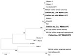
Figure. Phylogenetic tree based on the Leptospira flaB gene sequence. The sequences obtained in this study are indicated in boldface and have been deposited in DDBJ/GenBank/EMBL (accession numbers indicated). The sequences of...
A total of 134 plasma samples were collected from 93 patients. From 25 patients, 2–4 samples were collected; mean time from first to last collection was 6.1 (range 1–18) days. Among the 93 patients, 15 had a single MAT titer >400 and 4 showed a 4-fold increase in the MAT titer in paired samples. Most antibodies were against L. borgpetersenii serovar Tarassovi, followed by serovars Poi and Sejroe and L. interrogans serovars Losbanos and Manilae. PCR produced positive results for 4 patients, 2 of whom had a negative MAT titer. PCR-detected Leptospira strains were phylogenetically characterized by the flaB sequence (8,9); 3 were identical to those of isolates from rats in Metro Manila, but 1 was distinct (Figure).
Rapid-test results were available for 85 case-patients; 48 of 78 samples were positive by Leptospira Serology Kit and 5 of 7 were positive by ELISA. Of 27 cases tested by MAT and Leptospira Serology Kit, 6 were positive by both tests. One sample was negative by MAT and ELISA. Taken together, 149 (32%) of all cases underwent any diagnostic testing for leptospirosis, and results for 67 (45%) of these were laboratory confirmed. Clinical and laboratory findings of laboratory-confirmed and suspected cases, respectively, were almost identical except for the presence of jaundice (61.2% vs. 45.5%; p = 0.018), convulsions (4.5% vs. 0%; p = 0.003), and death (3% vs. 12.1%; p = 0.03). No plasma sample test results have been validated; thus, deduced incidence should be considered with caution. To explore the possibility of hantavirus co-infection contributing to the high CFRs, we screened samples for antibodies against hantavirus (10), but all were negative.
Risk factors for fatal leptospirosis were jaundice, anuria, and hemoptysis at admission. These are typical signs of the severe form of leptospirosis called Weil disease (11), confirming earlier work (12,13). Hemoptysis with high CFR (7 [47%] of 15) may have represented leptospirosis-associated severe pulmonary hemorrhagic syndrome (14). Some clinical features, including cough, seemed to be associated with lower risk for death, but minor symptoms in dying patients might have been overlooked. High leukocyte counts, blood urea nitrogen and creatinine levels, and lower platelet counts were also associated with death.
Deaths could be reduced if these indicators are detected and appropriate management introduced early. Laboratory data such as potassium level might also be helpful, but these tests are often not available for all suspected cases in resource-poor settings, especially during outbreaks. The trend of most patients being male is compatible with previous reports and thought to reflect higher exposure to contaminated water (6,12–14).
Although all patients received antimicrobial therapy, a considerable proportion died of acute respiratory distress syndrome and acute renal failure within only 2 days of admission, indicating that most patients sought care too late. The World Health Organization recommends starting antimicrobial therapy before the fifth day of disease onset (6), but for half of the case-patients reported here, it was started after the fifth day. Our results support the benefits of early initiation of antimicrobial therapy.
Renal failure was clearly a cause of death, but only 1 patient received peritoneal dialysis and another 7 were transferred to other hospitals for hemodialysis after only 3.4 days of hospitalization. This lack or delay of dialysis might have affected outcomes. San Lazaro Hospital is not sufficiently equipped for intensive care. Expanded access to peritoneal dialysis might reduce deaths from severe leptospirosis complicated by acute renal failure.
Leptospirosis is typically seen in resource-poor settings, where costly medical equipment such as dialyzers and ventilators are rarely accessible. For leptospirosis outbreak control and CFR reduction in leptospirosis-endemic regions, continuous case and environmental monitoring and early introduction of appropriate treatment for suspected cases are warranted.
Dr Amilasan is a senior resident physician at San Lazaro Hospital. His primary research interest is leptospirosis.
Acknowledgments
We thank Kenta Shimizu and Jiro Arikawa for testing for hantavirus, Maki Muto for testing for leptospirosis, and Mariko Saito for providing useful information in the field.
This work was supported by the Global Centers of Excellence Program, Institute of Tropical Medicine, Nagasaki University, Japan.
References
- Victoriano AF, Smythe LD, Gloriani-Barzaga N, Cavinta LL, Kasai T, Limpakarnjanarat K, Leptospirosis in the Asia Pacific region.BMC Infect Dis. 2009;9:147. DOIPubMedGoogle Scholar
- Yanagihara Y, Villanueva SY, Yoshida S, Okamoto Y, Masuzawa T. Current status of leptospirosis in Japan and Philippines.Comp Immunol Microbiol Infect Dis. 2007;30:399–413. DOIPubMedGoogle Scholar
- Pappas G, Papadimitriou P, Siozopoulou V, Christou L, Akritidis N. The globalization of leptospirosis: worldwide incidence trends.Int J Infect Dis. 2008;12:351–7. DOIPubMedGoogle Scholar
- Republic of the Philippines, National Disaster Coordinating Council. Situation report No. 52 on Tropical “Ondoy” (Ketsana) glide no. TC-2009–000205-PHL and Typhoon “Pepeng” (Parma) glide no. TC-2009–000214-PHL [cited 2011 Nov 4]. http://reliefweb.int/sites/reliefweb.int/files/resources/D87F541373D245BC4925767A000C5761-Full_Report.pdf
- Asian Disaster Reduction Center. Philippines’ country profile 2009 [cited 2011 Nov 4]. http://www.adrc.asia/countryreport/PHL/2009/PHL2009.pdf
- World Health Organization, International Leptospirosis Society. Human leptospirosis: guidance for diagnosis, surveillance and control. Geneva: The Organization; 2003.
- Barros AJ, Hirakata VN. Alternatives for logistic regression in cross-sectional studies: an empirical comparison of models that directly estimate the prevalence ratio.BMC Med Res Methodol. 2003;3:21. DOIPubMedGoogle Scholar
- Villanueva SY, Ezoe H, Baterna RA, Yanagihara Y, Muto M, Koizumi N, Serologic and molecular studies of Leptospira and leptospirosis among rats in the Philippines.Am J Trop Med Hyg. 2010;82:889–98. DOIPubMedGoogle Scholar
- Koizumi N, Muto M, Yamamoto S, Baba Y, Kudo M, Tamae Y, Investigation of reservoir animals of Leptospira in the northern part of Miyazaki Prefecture.Jpn J Infect Dis. 2008;61:465–8.PubMedGoogle Scholar
- Truong TT, Yoshimatsu K, Araki K, Lee BH, Nakamura I, Endo R, Molecular epidemiological and serological studies of hantavirus infection in northern Vietnam.J Vet Med Sci. 2009;71:1357–63. DOIPubMedGoogle Scholar
- Bharti AR, Nally JE, Ricaldi JN, Matthias MA, Diaz MM, Lovett MA, Leptospirosis: a zoonotic disease of global importance.Lancet Infect Dis. 2003;3:757–71. DOIPubMedGoogle Scholar
- Dupont H, Dupont-Perdrizet D, Perie JL, Zehner-Hansen S, Jarrige B, Daijardin JB. Leptospirosis: prognostic factors associated with mortality.Clin Infect Dis. 1997;25:720–4. DOIPubMedGoogle Scholar
- Panaphut T, Domrongkitchaiporn S, Thinkamrop B. Prognostic factors of death in leptospirosis: a prospective cohort study in Khon Kaen, Thailand.Int J Infect Dis. 2002;6:52–9. DOIPubMedGoogle Scholar
- Gouveia EL, Metcalfe J, de Carvalho AL, Aires TS, Villasboas-Bisneto JC, Queirroz A, Leptospirosis-associated severe pulmonary hemorrhagic syndrome, Salvador, Brazil.Emerg Infect Dis. 2008;14:505–8. DOIPubMedGoogle Scholar
Figure
Tables
Cite This Article1These authors contributed equally to this article.
Table of Contents – Volume 18, Number 1—January 2012
| EID Search Options |
|---|
|
|
|
|
|
|
Please use the form below to submit correspondence to the authors or contact them at the following address:
Motoi Suzuki, Department of Clinical Medicine, Institute of Tropical Medicine, Nagasaki University, Sakamoto 1-12-4, Nagasaki 852-8523, Japan
Top