Volume 18, Number 7—July 2012
Dispatch
Dobrava Hantavirus Infection Complicated by Panhypopituitarism, Istanbul, Turkey, 2010
Nevin Sarıgüzel1 , Jörg Hofmann1, Alper Tunga Canpolat, Ali Türk, Jakob Ettinger, Deniz Atmaca, Işın Akyar, Serap Yücel, Ender Arıkan, Yavuz Uyar, Dilek Y. Çağlayık, Ayşe Sesin Kocagöz, Ayşin Kaya, and Detlev H. Kruger
Figure 2
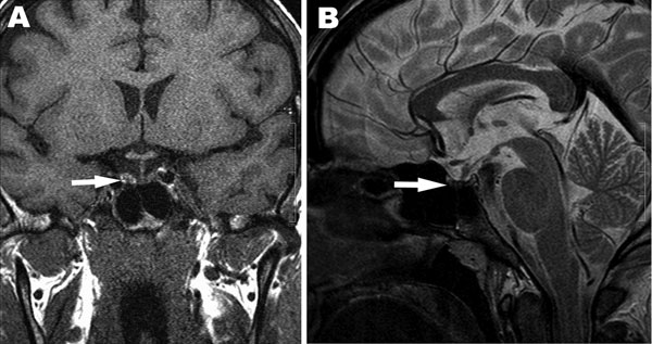
Figure 2. . . Magnetic resonance images showing hemorrhage of the pituitary gland and pituitary atrophy as indicated by arrows. A) T1-weighted coronal image shows high signal intensity on the right side of the adenohypophysis consistent with hemorrhage. B) T2-weighted sagittal image shows decreased pituitary gland height and heterogenous low signal intensity of the central adenohypophysis due to hemorrhagic infarction.
1These authors contributed equally to this article.
Page created: June 12, 2012
Page updated: June 12, 2012
Page reviewed: June 12, 2012
The conclusions, findings, and opinions expressed by authors contributing to this journal do not necessarily reflect the official position of the U.S. Department of Health and Human Services, the Public Health Service, the Centers for Disease Control and Prevention, or the authors' affiliated institutions. Use of trade names is for identification only and does not imply endorsement by any of the groups named above.