Identification of Cause of Posttransplant Cachexia by PCR
Joelle Guitard

, Sophie Edouard, Hubert Lepidi, Christine Segonds, Marion Grare, Marie-Laure Ranty-Quintyn, Isabelle Rouquette, Olivier Cointault, Lionel Rostaing, Nassim Kamar, and Florence Fenollar
Author affiliations: Centre Hospitalier Universitaire Rangueil, Toulouse, France (J. Guitard, C. Segonds, M. Grare, M.-L. Ranty-Quintyn, I. Rouquette, O. Cointault, L. Rostaing, N. Kamar); Université Aix-Marseille, Marseille, France (S. Edouard, H. Lepidi, F. Fenollar); Pôle de Maladies Infectieuses, Marseille (S. Edouard, F. Fenollar); and Université Paul Sabatier, Toulouse (L. Rostaing, N. Kamar)
Main Article
Figure
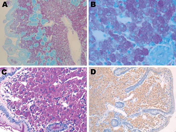
Figure. . . Duodenal biopsy specimen from the patient with posttransplant cachexia. Ziehl–Neelsen acid staining of a patient biopsy specimen, showing partially reduced villous architecture at low magnification, with numerous Ziehl–Neelsen-positive macrophages within the lamina propria (A, original magnification ×50). High magnification clearly demonstrates mycobacteria as bacillary particles in the macrophage cytoplasm (B, original magnification ×400). C) Macrophages within the lamina propria are periodic acid-Schiff–positive, diastase-resistant particles but do not show a morphologic granular pattern (original magnification ×200). D) Immunohistochemical staining with a polyclonal rabbit antibody against Tropheryma whipplei shows low immunostaining without a granular pattern (antibody used at a dilution of 1:2,000, hemalun counterstain, original magnification ×100).
Main Article
Page created: July 18, 2012
Page updated: July 18, 2012
Page reviewed: July 18, 2012
The conclusions, findings, and opinions expressed by authors contributing to this journal do not necessarily reflect the official position of the U.S. Department of Health and Human Services, the Public Health Service, the Centers for Disease Control and Prevention, or the authors' affiliated institutions. Use of trade names is for identification only and does not imply endorsement by any of the groups named above.