Volume 19, Number 11—November 2013
Research
Atypical Scrapie Prions from Sheep and Lack of Disease in Transgenic Mice Overexpressing Human Prion Protein
Figure 2
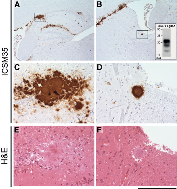
Figure 2. . Immunohistochemical analysis of cattle bovine spongiform encephalopathy (BSE) prion–infected 129MM Tg35c mouse brain. Hippocampal region (A) and striatum (B) from a transgenic 129MM Tg35c mouse with subclinical prion infection culled 700 days after inoculation with cattle BSE prion inoculum I038. Panels A–D show abnormal prion protein (PrP) immunoreactivity stained with monoclonal antibody ICSM35 against PrP. Panels E and F show hematoxylin and eosin–stained sections. Boxed regions in panels A and B are shown at higher power magnification in panels C and E, and D and F, respectively. The inset in panel B shows an immunoblot in which monoclonal antibody 3F4 against PrP was used, which demonstrates type 4 PrPSc in 10 μL of PK-digested 10% (w/v) brain homogenate prepared from the contralateral side of the same brain. Scale bar indicates 1.2 mm for panels A and B, 160 μm for panels C–F.