Volume 19, Number 4—April 2013
Research
Occult Hepatitis B Virus Infection in Chacma Baboons, South Africa
Figure 5
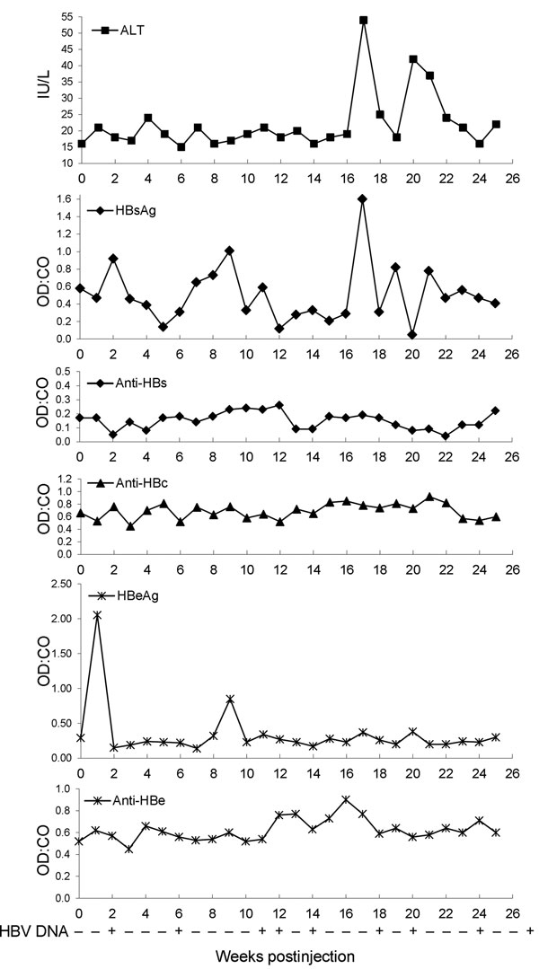
Figure 5. . . Levels of alanine aminotransferase (ALT) and hepatitis B virus (HBV) serologic markers and detection of HBV DNA in baboon 2. Serum obtained from baboon 2 (which was injected with serum from baboon 9732), and ALT and HBV serologic marker levels were measured at weekly intervals after injection. Serum for week 0 levels was obtained just before injection. OD:CO indicates optical density:cutoff value ratios. An OD:CO >1 indicates a positive result. HBV DNA was detected by nested PCR amplification (255–761 in the first round and 459–710 in the second round) of a 252-bp region of the virus surface gene by using DNA extracted from serum obtained from the infected baboon at weekly intervals. HBsAg, HBV surface antigen; Anti-HBs, antibody against HBV surface antigen; Anti-HBc, antibody against HBV core antigen; HBeAg, HBV e antigen; Anti-HBe, antibody against HBV e antigen. + indicates that the sample amplified successfully at this time point, and – indicates no amplification of virus DNA. The week 26 time point indicates the result of amplification by using DNA extracted from serum at necropsy, and the final time point indicates successful amplification of the virus product from DNA extracted from liver tissue obtained from baboon 2 at necropsy.