Foodborne Transmission of Bovine Spongiform Encephalopathy to Nonhuman Primates
Edgar Holznagel

, Barbara Yutzy, Walter Schulz-Schaeffer, Carina Kruip, Uwe Hahmann, Pär Bierke, Juan-Maria Torres, Yong-Sun Kim, Achim Thomzig, Michael Beekes, Gerhard Hunsmann, and Johannes Loewer
Author affiliations: Paul-Ehrlich-Institut, Langen, Germany (E. Holznagel, B. Yutzy, C. Kruip, J. Loewer); University of Göttingen, Göttingen, Germany (W. Schulz-Schaeffer); German Primate Centre, Göttingen (U. Hahmann, G. Hunsmann); Swedish Institute for Infectious Disease Control, Solna, Sweden (P. Bierke); Centro de Investigación en Sanidad Animal, Madrid, Spain (J.-M. Torres); Hallym University, Anyang, Gyeonggi-Do, South Korea (Y.-S. Kim); Robert-Koch-Institut, Berlin, Germany (A. Thomzig, M. Beekes)
Main Article
Figure 3
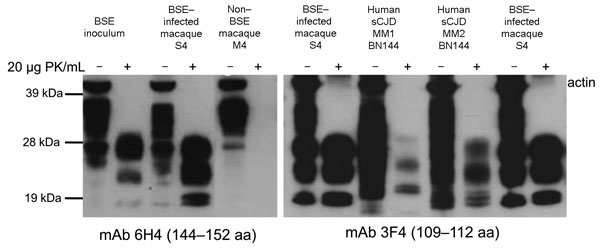
Figure 3. . Western immunoblot analysis of proteinease-resistant prion protein (PrPres) (from brain). The size of the nonglycosylated band was either 21 kDa (termed type 1, sCJD case BN141) or 19 kDa (termed type 2, sCJD case BN144). Diglycosylated PrPres predominated in the BSE inoculum and in all macaques showing neurologic signs (macaque S4 is shown as a representative example for all symptomatic cases), and the pattern is termed type 2B to distinguish it from type 2 cases in which the monoglycosylated form predominated (Type 2A, sCJD case BN144). Two different mAbs were used for routine immunodetection: 6H4 (left panel) and 3F4 (right panel). Blots were co-stained with a polyclonal anti-actin antiserum. BSE, bovine spongiform encephalopathy; sCJD, sporadic Creutzfeldt-Jakob disease; PK, proteinase K; mAb, monoclonal antibody.
Main Article
Page created: April 24, 2013
Page updated: April 24, 2013
Page reviewed: April 24, 2013
The conclusions, findings, and opinions expressed by authors contributing to this journal do not necessarily reflect the official position of the U.S. Department of Health and Human Services, the Public Health Service, the Centers for Disease Control and Prevention, or the authors' affiliated institutions. Use of trade names is for identification only and does not imply endorsement by any of the groups named above.