Volume 19, Number 7—July 2013
Dispatch
Clinical Findings for Early Human Cases of Influenza A(H7N9) Virus Infection, Shanghai, China
Shuihua Lu1 , Yufang Zheng1, Tao Li1, Yunwen Hu1, Xinian Liu, Xiuhong Xi, Qingguo Chen, Qingle Wang, Ye Cao, Yanbing Wang, Lijun Zhou, Douglas Lowrie, and Jing Bao
Figure 1
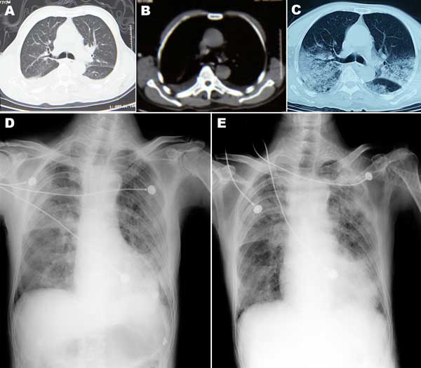
Figure 1. . . Chest computed tomography (CT) scan and radiograph images of patient (case-patient 1) in a study of 4 persons with early cases of influenza A(H7N9) virus infection, Shanghai, China. Images were taken 1, 5, 7, and 11 days after illness onset. A, B) CT scan images on day 1, showing bilateral pleural effusion but no obvious lesions. C) CT scan image on day 5, showing extensive ground-glass opacity and consolidation. D, E) x-ray images on days 7 and 11, respectively, showing reduced light transmittance on both sides of the lung.
1These authors contributed equally to this article.
Page created: June 18, 2013
Page updated: June 18, 2013
Page reviewed: June 18, 2013
The conclusions, findings, and opinions expressed by authors contributing to this journal do not necessarily reflect the official position of the U.S. Department of Health and Human Services, the Public Health Service, the Centers for Disease Control and Prevention, or the authors' affiliated institutions. Use of trade names is for identification only and does not imply endorsement by any of the groups named above.