Volume 20, Number 1—January 2014
Dispatch
Pathogenic Pseudorabies Virus, China, 2012
Xiuling Yu1, Zhi Zhou1, Dongmei Hu1, Qian Zhang1, Tao Han, Xiaoxia Li, Xiaoxue Gu, Lin Yuan, Shuo Zhang, Baoyue Wang, Ping Qu, Jinhua Liu, Xinyan Zhai, and Kegong Tian
Figure 1
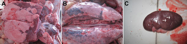
Figure 1. . Necropsy specimens from pigs infected with pseudorabies virus. A) Pulmonary consolidation in the lung. B) Edema and hemorrhage of lung. C) Kidney with many yellow-white necrotic spots (arrows).
1These authors contributed equally to this article.
Page created: January 03, 2014
Page updated: January 03, 2014
Page reviewed: January 03, 2014
The conclusions, findings, and opinions expressed by authors contributing to this journal do not necessarily reflect the official position of the U.S. Department of Health and Human Services, the Public Health Service, the Centers for Disease Control and Prevention, or the authors' affiliated institutions. Use of trade names is for identification only and does not imply endorsement by any of the groups named above.