Volume 20, Number 10—October 2014
Letter
Marburgvirus Resurgence in Kitaka Mine Bat Population after Extermination Attempts, Uganda
Figure
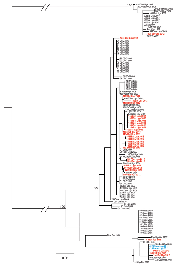
Figure. Phylogeny of concatenated marburgvirus nucleoprotein (NP) and viral protein 35 (VP35) gene fragments as determined by using the maximum-likelihood method. Sequences from the NP (289–372 nt) and VP35 (203–213 nt) genes were amplified and determined from viral RNA and then sequenced as described elsewhere (4). Sequence names shown in red font represent those generated from samples collected from bats during the November 2012 outbreak investigation at Kitaka Mine, Uganda. Sequence names in blue font represent those generated from samples obtained from marburgvirus-infected persons in Kabale and Ibanda, Uganda, in 2012. Multiple sequence alignments were generated, and a maximum-likelihood analysis was conducted on concatenated NP and VP35 (208–580 nt) sequences by using the PhyML method in conjunction with the GTR+I+G nucleotide substitution model implemented in SeaView version 4.2.12 (10). NP and VP35 gene sequences determined from samples in this study (in red) were submitted to GenBank (accession nos. KJ747211–KJ747234 and KJ747235–KJ747253, respectively). Bayesian posterior probabilities above 50 are shown at the nodes. Scale bar indicates nucleotide substitutions per site. Ang, Angola; DRC, Democratic Republic of Congo; Gab, Gabon; Ger, Germany; Ken, Kenya; Net, Netherlands; Rav, Ravn virus; Uga, Uganda; Zim, Zimbabwe.
References
- Adjemian J, Farnon EC, Tschioko F, Wamala JF, Byaruhanga E, Bwire GS, Outbreak of Marburg hemorrhagic fever among miners in Kamwenge and Ibanda Districts, Uganda, 2007. J Infect Dis. 2011;204(Suppl 3):S796–9 . DOIPubMedGoogle Scholar
- Timen A, Koopmans MP, Vossen AC, van Doornum GJ, Gunther S, van den Berkmortel F, Response to imported case of Marburg hemorrhagic fever, the Netherlands. Emerg Infect Dis. 2009;15:1171–5 . DOIPubMedGoogle Scholar
- Centers for Disease Control and Prevention. Imported case of Marburg hemorrhagic fever–Colorado, 2008. MMWR Morb Mortal Wkly Rep. 2009;58:1377–81 .PubMedGoogle Scholar
- Amman BR, Carroll SA, Reed ZD, Sealy TK, Balinandi S, Swanepoel R, Seasonal pulses of Marburg virus circulation in juvenile Rousettus aegyptiacus bats coincide with periods of increased risk of human infection. PLoS Pathog. 2012;8:e1002877 . DOIPubMedGoogle Scholar
- Towner JS, Amman BR, Sealy TK, Carroll SA, Comer JA, Kemp A, Isolation of genetically diverse Marburg viruses from Egyptian fruit bats. PLoS Pathog. 2009;5:e1000536 . DOIPubMedGoogle Scholar
- Donnelly CA, Woodroffe R, Cox DR, Bourne J, Gettinby G, Le Fevre AM, Impact of localized badger culling on tuberculosis incidence in British cattle. Nature. 2003;426:834–7. DOIPubMedGoogle Scholar
- Swanepoel R. Rabies. In: Coetzer JAW, Tustin RC, editors. Infectious diseases of livestock. 2nd ed. Cape Town (South Africa): Oxford University Press Southern Africa; 2014. p. 1123–82.
- Albariño CG, Shoemaker T, Khristova ML, Wamala JF, Muyembe JJ, Balinandi S, Genomic analysis of filoviruses associated with four viral hemorrhagic fever outbreaks in Uganda and the Democratic Republic of the Congo in 2012. Virology. 2013;442:97–100. DOIPubMedGoogle Scholar
- Maganga GD, Bourgarel M, Ella GE, Drexler JF, Gonzalez JP, Drosten C, Is Marburg virus enzootic in Gabon? J Infect Dis. 2011;204(Suppl 3):S800–3 . DOIPubMedGoogle Scholar
- Gouy M, Guindon S, Gascuel O. SeaView version 4: a multiplatform graphical user interface for sequence alignment and phylogenetic tree building. Mol Biol Evol. 2010;27:221–4. DOIPubMedGoogle Scholar